 plumi
Cell structure: OCR A‑Level Biology A questions
Every OCR A-Level Biology A exam question on 2.1.1 Cell structure, 2016–2025, with its mark scheme and examiner’s report.
Find questions1,984
Calculations
Q16(a)(i)
The whole paper ↗
Q16(a)(ii)
The whole paper ↗
Q16(a)(iii)
The whole paper ↗

Q22(b)(i)
The whole paper ↗
Q23(a)(ii)
The whole paper ↗
Q26(b)
The whole paper ↗
Q21(a)
The whole paper ↗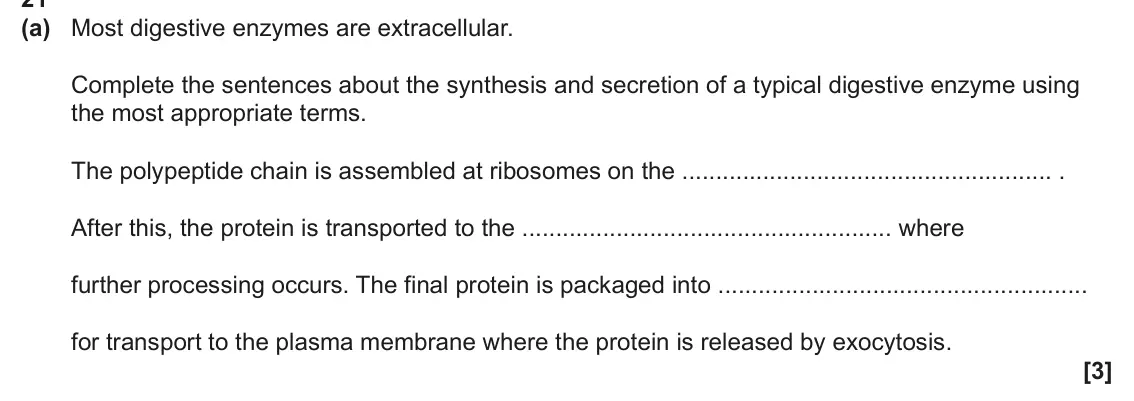


Q17(b)(iii)
The whole paper ↗
Q1(a)(i)
The whole paper ↗
Q1(a)(ii)
The whole paper ↗
Q3(b)(ii)
The whole paper ↗

Q5(a)(i)
The whole paper ↗
Q5(a)(ii)
The whole paper ↗
Q5(b)
The whole paper ↗
Q5(c)(i)
The whole paper ↗
All 97 questions on Cell structure, as a list
- June 2025, Paper 2, Q16(a)(i) (2 marks): Identify two pieces of evidence that suggest that the image was produced by a light microscope rather than a scanning electron microscope. 1 2 [2]
- June 2025, Paper 2, Q16(a)(ii) (2 marks): Calculate the horizontal diameter of the cell shown in the image. Give your answer in μm. Diameter = μm [2]
- June 2025, Paper 2, Q16(a)(iii) (2 marks): Explain why staining is useful in microscopy. Use structures visible in the image to support your answer. [2]
- June 2025, AS Paper 1, Q1 (1 mark): Which statement correctly describes microscopes? A Light microscopes can produce higher resolution images than electron microscopes. B Scanning electron microsc
- June 2025, AS Paper 1, Q22(b)(i) (1 mark): The DNA in the nucleus of a eukaryotic cell is associated with histone proteins. State one other feature of DNA in a eukaryotic cell that is different from DNA
- June 2025, AS Paper 1, Q23(a)(ii) (2 marks): Calculate the magnification of the B lymphocyte. Give your answer to 2 significant figures. Magnification = [2]
- June 2025, AS Paper 1, Q26(b) (2 marks): A student is studying erythrocytes and neutrophils in a blood sample. They smear a drop of the blood sample across a microscope slide and place it under a light
- June 2024, Paper 1, Q21(a) (3 marks): Most digestive enzymes are extracellular. Complete the sentences about the synthesis and secretion of a typical digestive enzyme using the most appropriate term
- June 2024, Paper 2, Q4 (1 mark): Microscopes vary in terms of magnification and resolution. Which option describes the terms magnification and resolution? A Magnification is the size of an imag
- June 2024, Paper 2, Q14 (1 mark): Identify the structure labelled X. A Centriole B Lysosome C Mitochondrion D Nucleolus Your answer [1]
- June 2024, Paper 2, Q15 (1 mark): Identify the structure labelled Y. A Cell wall B Cilium C Plasma membrane D Smooth ER Your answer [1]
- June 2024, Paper 2, Q17(b)(iii) (3 marks): The image was taken using a × 40 objective lens. The eyepiece graticule scale for the × 40 objective lens was calibrated using the stage micrometer shown below.
- June 2024, Paper 3, Q1(a)(i) (1 mark): Name the structure labelled A. A [1]
- June 2024, Paper 3, Q1(a)(ii) (1 mark): Based on your knowledge, estimate the diameter of the E. coli cell. Give your answer in µm. Diameter = µm [1]
- June 2024, Paper 3, Q3(b)(ii) (2 marks): This is a light micrograph of a transverse section through the stem of P. vulgaris. V × × 90 The diameter of one of the vascular bundles is shown by the line la
- June 2024, AS Paper 1, Q8 (1 mark): Bacillus thuringiensis (Bt) is a species of bacterium that lives in soil. Bt makes proteins that are toxic to some insects when eaten. Which process does not oc
- June 2024, AS Paper 2, Q5(a)(i) (2 marks): Some students collect a sample of pond water to observe under a light microscope. They pour some of the pond water onto a microscope slide. Describe how they co
- June 2024, AS Paper 2, Q5(a)(ii) (2 marks): Explain how the students would use the different objective lenses of a light microscope to focus and observe the pond water sample at high power magnification.
- June 2024, AS Paper 2, Q5(b) (2 marks): Fig. 5.1 shows a photomicrograph of human cheek cells observed by the students under a light microscope. Fig. 5.1 Magnification ×400 Fig. 5.2 shows a drawing ma
- June 2024, AS Paper 2, Q5(c)(i) (2 marks): Fig. 5.3 shows a photomicrograph of a liver cell taken from a transmission electron microscope (TEM). Fig. 5.3 Nuclear pore Nuclear envelope Vesicle Golgi appar
- June 2024, AS Paper 2, Q5(c)(ii) (4 marks): Describe how the rough endoplasmic reticulum and the Golgi apparatus are involved in the production of a secretory vesicle that contains protein. [4]
- June 2024, AS Paper 2, Q5(d) (3 marks): Outline the importance of the cytoskeleton. [3]
- June 2023, Paper 1, Q18(b)(i) (3 marks): Give three functions of the cytoskeleton. 1 2 3 [3]
- June 2023, Paper 1, Q18(b)(ii) (2 marks): Suggest how a mutation in cytoskeleton genes could cause a disease of the nervous system. [2]
- June 2023, Paper 2, Q5 (1 mark): A student was observing and measuring cells using a light microscope. Which option describes a method that would enable the student to accurately measure the le
- June 2023, Paper 2, Q6 (1 mark): Which of the following describes, from lowest to highest, the resolution of images than can be achieved by different types of microscope? A Light microscope sca
- June 2023, AS Paper 1, Q7 (1 mark): A student observes xylem tissue under a microscope. The student uses an eyepiece graticule and a stage micrometer to measure the diameter of a xylem vessel. Eye
- June 2023, AS Paper 1, Q8 (1 mark): What is the pathway taken by a protein that has been made and is to be secreted from a cell? A Ribosome vesicle Golgi apparatus cytoplasm B Ribosome vesicle rou
- June 2023, AS Paper 1, Q9 (1 mark): What are the roles of the cytoskeleton in a human skin cell? A Movement of the cell through its environment and providing mechanical strength to the cell B Move
- June 2023, AS Paper 1, Q10 (1 mark): Which feature is associated with a prokaryotic cell? A Cell walls made of chitin B Extrachromosomal DNA that is circular C Linear DNA that can form plasmids D 7
- June 2023, AS Paper 1, Q28(a) (4 marks): Four organelles are listed in the table below. Complete the table. For each organelle place a tick (3) in the box for a correct description and leave the box em
- June 2022, Paper 3, Q2(a)(iii) (2 marks): The diameter of another blood cell is represented by the line W in Fig. 2.1. The magnification used to produce Fig. 2.1 was × 800. Calculate the actual diameter
- June 2022, Paper 3, Q2(b)(ii) (2 marks): A scientist calculated two values for the lysosomes in a white blood cell: mean volume of a lysosome = 6.5 × 10⁻¹⁴ cm3 • mean number of H+ ions per lysosome = 1
- June 2022, Paper 3, Q2(b)(iii) (1 mark): The formula used to calculate pH is pH = -log [H+] where [H+] is H+ ion concentration in mol dm–3. Use your answer from part (ii) to calculate the mean pH of th
- June 2022, Paper 3, Q2(b)(iv) (1 mark): The scientist stained the lysosomes in a sample of living white blood cells. The table shows the properties of five stains, A to E. Stain Properties Suitable to
- June 2022, Paper 3, Q2(c) (2 marks): Differential staining can be used to distinguish between bacteria with thick cell walls and bacteria with thin cell walls. Four substances are used when differe
- June 2022, AS Paper 1, Q18 (1 mark): A student is studying three unicellular organisms: the bacterium Escherichia coli, the protoctist Euglena gracilis and the fungus Saccharomyces cerevisiae. Whic
- June 2022, AS Paper 1, Q25(b)(ii) (3 marks): Name the type of microscope used to produce the photomicrograph and explain the reasons for your choice. Name of microscope Reasons for your choice [3]
- June 2022, AS Paper 2, Q4(a)(ii) (2 marks): The magnification of the microscope used to observe the cells in Fig. 4.1 was ×950. Calculate the diameter of cell X in Fig. 4.1. Give your answer in micrometre
- November 2021, Paper 1, Q3 (1 mark): Which of the options describes the path taken by proteins, such as digestive enzymes, that are exported from a cell? A Golgi apparatus rough endoplasmic reticul
- November 2021, AS Paper 1, Q7 (1 mark): Which of the stains, A to D, would be chosen to bind to the phosphate group of DNA to make chromosomes more visible when using a light microscope? A carbolfuchs
- November 2021, AS Paper 1, Q21(b)(ii) (3 marks): Tubulin is a globular protein that can polymerise to form the cell cytoskeleton. One example of this is the formation of microtubules, which form the spindle fi
- November 2021, AS Paper 1, Q21(b)(iii) (2 marks): Suggest two ways tubulin is essential to protein synthesis and protein secretion in eukaryotic cells. 1 2 [2]
- November 2021, AS Paper 1, Q22(a) (4 marks): Explain how to measure the diameter of the nucleus of one of the white blood cells, when observing the cells through a light microscope. [4]
- November 2021, AS Paper 1, Q22(b)(i) (2 marks): Complete the table below about microscopes and their images. Laser scanning Scanning electron Transmission confocal microscope microscope electron microscope Ma
- November 2021, AS Paper 1, Q24(b)(i) (1 mark): Suggest one feature of a bacterium the phazolicin might attack. [1]
- November 2020, Paper 1, Q1 (1 mark): Which of the following statements, A to D, about differential staining is not true? A Differential staining can distinguish between different organelles. B Diff
- November 2020, Paper 1, Q2 (1 mark): Which of the following statements, A to D, about microscopes is correct? A A light microscope with an eyepiece lens magnification of × 10 and an objective lens
- November 2020, Paper 1, Q18(b)(i) (1 mark): Explain one advantage of using a confocal microscope in this study. [1]
- November 2020, Paper 1, Q19(a)(i) (1 mark): State the type of microscope used to produce this image. [1]
- November 2020, Paper 1, Q19(c)(i) (2 marks): Give two structural features of mitochondria that support this theory. 1 2 [2]
- November 2020, Paper 1, Q20(c)(ii) (2 marks): A light microscope can be used to observe yeast cells. State the equipment that would be needed, in addition to a microscope, to measure the average diameter of
- November 2020, Paper 2, Q5 (1 mark): The cell labelled E shows a parasite called Trypanosoma. Which of the following statements is/are evidence that Trypanosoma is a eukaryote? a nucleus is present
- November 2020, Paper 2, Q19(c)(iii) (2 marks): A diagram of a moss sperm is shown in Fig. 19.3. flagella nucleus Fig. 19.3 The flagella allow the sperm to move towards an egg. Suggest and explain another ada
- November 2020, Paper 3, Q4(a)(i) (1 mark): Suggest the function of Protein A. [1]
- November 2020, Paper 3, Q4(a)(ii) (1 mark): Suggest the function of Protein C. [1]
- November 2020, Paper 3, Q4(a)(iii) (1 mark): An antibiotic called A22 binds irreversibly to Protein B. Despite its antibiotic properties, A22 is not used in humans. Suggest why scientists have advised that
- November 2020, AS Paper 1, Q23(a) (3 marks): Describe the structure of the rough endoplasmic reticulum. [3]
- November 2020, AS Paper 1, Q26(a)(iii) (1 mark): Describe how the genetic material is arranged in organisms in the same kingdom as Y. pestis. [1]
- November 2020, AS Paper 2, Q1(a)(i) (3 marks): Identify one incorrect label and explain your answer. Incorrect label Explanation [3]
- November 2020, AS Paper 2, Q1(a)(ii) (3 marks): State three changes, other than to the labels, to Fig. 1.2 that the student would need to make to improve the biological drawing. 1 2 3 [3]
- November 2020, AS Paper 2, Q1(b) (4 marks): Both a transmission electron microscope (TEM) and a scanning electron microscope (SEM) can be used to view the same cell. However, the images formed will be dif
- November 2020, AS Paper 2, Q2(a)(i) (1 mark): Suggest why goblet cells have large numbers of the cellular component labelled A. [1]
- November 2020, AS Paper 2, Q2(a)(ii) (2 marks): Suggest how the role of the cellular component labelled B is relevant to the function of the goblet cell. [2]
- November 2020, AS Paper 2, Q2(b)(i) (2 marks): Mucus is present in goblet cells as condensed granules. Some studies reveal that when secreted, the mucus expands to 500 times its volume in 20 ms. 40 cm3 of mu
- June 2019, Paper 1, Q16(c) (1 mark): Brown fat is a type of tissue. Brown fat has a higher need for oxygen because fat cells in this tissue carry out aerobic respiration at a higher rate than fat c
- June 2019, Paper 3, Q1(a)(i) (2 marks): Identify the cellular components shown at A and B. A B [2]
- June 2019, Paper 3, Q1(a)(ii) (2 marks): The real size of the line between C and D on Fig. 1.1 is 1.4 × 10⁻⁶ m. Calculate the magnification that was used to produce the image in Fig. 1.1. Give your ans
- June 2019, Paper 3, Q1(a)(iii) (2 marks): Fig. 1.2 shows a student’s drawing of another sago pondweed cell, which was observed under a light microscope. The student used a sharp pencil but did not label
- June 2019, Paper 3, Q1(a)(iv) (2 marks): The student stained a sago pondweed sample to improve the contrast between cellular components when viewed under a microscope. The student used the following pr
- June 2019, AS Paper 1, Q25(b)(i) (1 mark): Name the molecule that is found in the cell walls of bacterial cells. [1]
- June 2019, AS Paper 2, Q2(a) (2 marks): Calculate the image length, in µm, represented by the scale bar. Give your answer to 2 significant figures. length = µm [2]
- June 2019, AS Paper 2, Q2(c)(i) (3 marks): Explain why it is important to use a differential stain when examining a blood smear under the microscope. [3]
- June 2019, AS Paper 2, Q2(c)(ii) (4 marks): Using Fig. 2 on the insert, and the information provided, suggest and explain why the cytoplasm of cell C and cell D reacted differently to the stain. [4]
- June 2018, Paper 2, Q1 (1 mark): Which of the following best describes a microscope with high resolution? A The microscope can distinguish structures that are very close together. B The microsc
- June 2018, Paper 2, Q2 (1 mark): The image below shows a tardigrade, Echiniscus granulatus, viewed from the underneath. The magnification is × 110. How long is the tardigrade in real life? A 11
- June 2018, Paper 2, Q16(b)(i) (3 marks): The volume of cell A is 5.4 × 104μm3. Assume that cell B is spherical. Calculate the volume of cell B. Use the formula: volume of sphere = 4 3πr3 Give your answ
- June 2018, Paper 2, Q16(b)(ii) (2 marks): State the type of microscope that was used to view these images. Justify your answer. [2]
- June 2018, Paper 3, Q1(a) (2 marks): The table below contains statements about the root cells of an onion. Place ticks (3) in the boxes in the table to indicate whether the statements are true or f
- June 2018, AS Paper 1, Q1 (1 mark): Which organelle, A to D, is not involved in the production and secretion of enzymes in eukaryotes? A golgi apparatus B ribosomes C smooth endoplasmic reticulum
- June 2018, AS Paper 1, Q12 (1 mark): The bacterium Sorangium cellulosum and the fungus Armillaria mellea are both found in soil. Which of the rows, A to D, correctly shows the structures present in
- June 2018, AS Paper 1, Q23(a) (2 marks): Calculate the magnification of this image. [2]
- June 2018, AS Paper 2, Q3(a) (1 mark): A cytoskeleton is present in all eukaryotic cells. One of its functions is to control the movement of organelles. State how the cytoskeleton moves organelles ar
- June 2018, AS Paper 2, Q3(b) (4 marks): Epithelial cells in the airways of mammals play an essential role in defences against pathogens. Explain the function of epithelial cells in the airways of mamm
- June 2017, Paper 1, Q3 (1 mark): A student counted stomata on a leaf using a light microscope. The image below shows the stomata that were visible. The image magnification is ×60. Which of the
- June 2017, Paper 1, Q4 (1 mark): Which of the options, A to D, occurs in the nucleus of a cell? A synthesis of enzymes B synthesis of RNA C modification of polypeptides D synthesis of carbohydr
- June 2017, Paper 1, Q17(b)(i) (3 marks): The diameter of the Kupffer cell in the image is 9.1 cm. Assuming it is spherical, calculate the actual volume of this cell. Give your answer to four significan
- June 2017, Paper 1, Q17(b)(ii) (2 marks): Which type of microscope has been used to obtain this image? Explain your answer. [2]
- June 2017, Paper 1, Q20(e) (1 mark): Anaerobic respiration in yeast cells requires enzymes. Which organelle is responsible for synthesising these enzymes? [1]
- June 2017, AS Paper 1, Q21(a)(ii) (1 mark): Suggest a stain that the student could have used to highlight the nuclei of these cells. [1]
- June 2017, AS Paper 1, Q21(b) (2 marks): The student used an eyepiece graticule, which is shown in Fig. 21. The student calibrated the graticule before carrying out the root tip squash. He observed tha
- June 2017, AS Paper 1, Q22(b) (3 marks): Humans use the enzyme α-amylase to break down polysaccharides in food for absorption into the blood. The gene for human α-amylase is found on chromosome 1. The
- June 2017, AS Paper 2, Q1(a)(i) (1 mark): Cell B in Fig. 1.1 was observed using an electron microscope. Give one piece of evidence from Fig. 1.1 that supports this. [1]
- June 2017, AS Paper 2, Q1(a)(ii) (1 mark): Give one way that an image produced by a laser scanning confocal microscope differs from that produced by an electron microscope. [1]
- June 2016, AS Paper 1, Q1 (1 mark): Microscopes vary in their magnification and resolution. Which of the rows, A to D, in the table below is correct? Transmission electron Scanning electron Light
- June 2016, AS Paper 1, Q6 (1 mark): Which of the following structures, A to D, are found in prokaryotes and in eukaryotes? A a cell wall made of peptidoglycan B circular genomic DNA C a nucleus su
- June 2016, AS Paper 1, Q21(b)(i) (3 marks): Identify the parts of the cell labelled X, Y and Z. X Y Z [3]
Questions, mark schemes and examiner’s reports are © OCR, from their published past papers. Plumi isn’t affiliated with any exam board. Want whole papers? Every OCR A-Level Biology A paper, or our free OCR-style practice pack.
Rather write your answers by hand on iPad? Plumi marks them against the same mark schemes.