 plumi
Magnification and resolution: OCR A‑Level Biology A questions
OCR A-Level Biology A past-paper questions on 2.1.1.2 Magnification and resolution, 2016–2025, with mark schemes and examiner’s reports where available.
Paper
All papers
Exam
All years
Calculations
Q16(a)(ii)
The whole paper ↗
- Your answer
Q23(a)(ii)
The whole paper ↗- Your answer
Q17(b)(iii)
The whole paper ↗
Q3(b)(ii)
The whole paper ↗
Q5(c)(i)
The whole paper ↗- Your answer
- Your answer
- Your answer
Q2(a)(iii)
The whole paper ↗Q25(b)(ii)
The whole paper ↗Q4(a)(ii)
The whole paper ↗Q22(a)
The whole paper ↗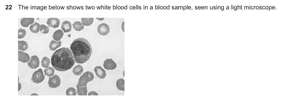
Q22(b)(i)
The whole paper ↗- Your answer
Q20(c)(ii)
The whole paper ↗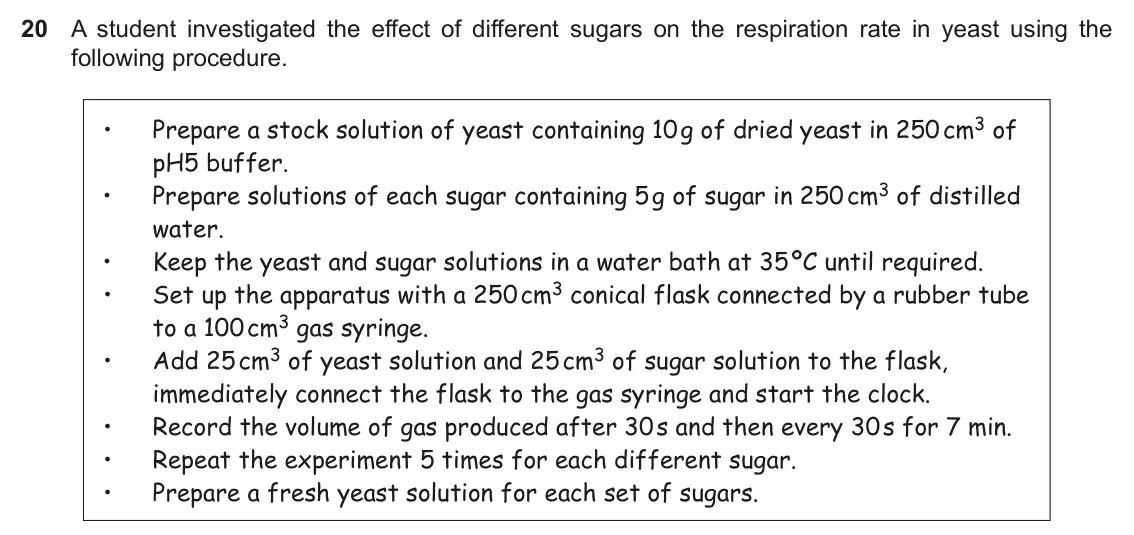
Q1(b)
The whole paper ↗Q1(a)(ii)
The whole paper ↗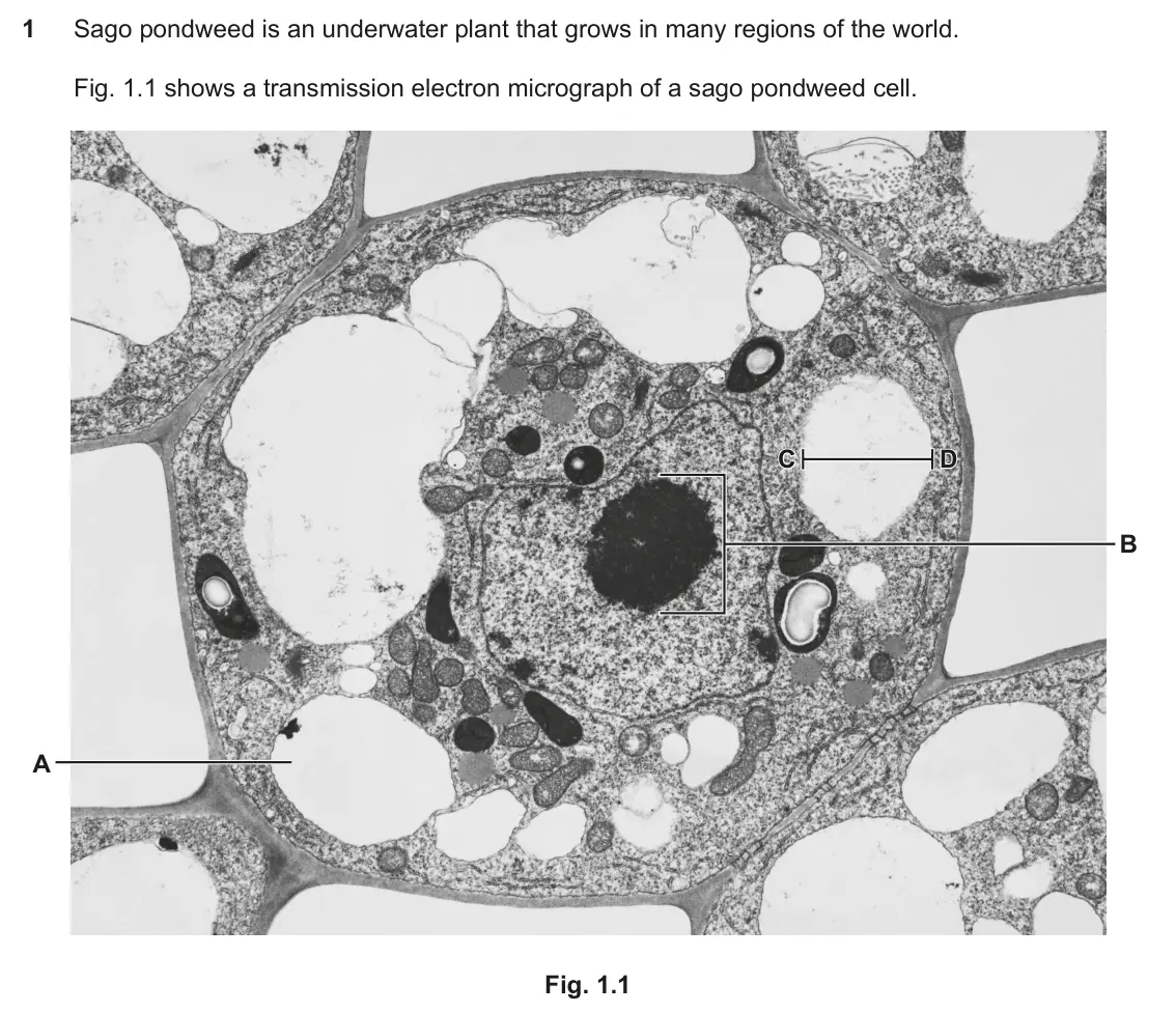
Q2(a)
The whole paper ↗
All 31 questions on Magnification and resolution, as a list
- June 2025, Paper 2, Q16(a)(ii) (2 marks): Calculate the horizontal diameter of the cell shown in the image. Give your answer in μm. Diameter = μm [2]
- June 2025, AS Paper 1, Q1 (1 mark): Which statement correctly describes microscopes? A Light microscopes can produce higher resolution images than electron microscopes. B Scanning electron microsc
- June 2025, AS Paper 1, Q23(a)(ii) (2 marks): Calculate the magnification of the B lymphocyte. Give your answer to 2 significant figures. Magnification = [2]
- June 2024, Paper 2, Q4 (1 mark): Microscopes vary in terms of magnification and resolution. Which option describes the terms magnification and resolution? A Magnification is the size of an imag
- June 2024, Paper 2, Q17(b)(iii) (3 marks): The image was taken using a × 40 objective lens. The eyepiece graticule scale for the × 40 objective lens was calibrated using the stage micrometer shown below.
- June 2024, Paper 3, Q3(b)(ii) (2 marks): This is a light micrograph of a transverse section through the stem of P. vulgaris. V × × 90 The diameter of one of the vascular bundles is shown by the line la
- June 2024, AS Paper 2, Q5(c)(i) (2 marks): Fig. 5.3 shows a photomicrograph of a liver cell taken from a transmission electron microscope (TEM). Fig. 5.3 Nuclear pore Nuclear envelope Vesicle Golgi appar
- June 2023, Paper 2, Q5 (1 mark): A student was observing and measuring cells using a light microscope. Which option describes a method that would enable the student to accurately measure the le
- June 2023, Paper 2, Q6 (1 mark): Which of the following describes, from lowest to highest, the resolution of images than can be achieved by different types of microscope? A Light microscope sca
- June 2023, AS Paper 1, Q7 (1 mark): A student observes xylem tissue under a microscope. The student uses an eyepiece graticule and a stage micrometer to measure the diameter of a xylem vessel. Eye
- June 2022, Paper 3, Q2(a)(iii) (2 marks): The diameter of another blood cell is represented by the line W in Fig. 2.1. The magnification used to produce Fig. 2.1 was × 800. Calculate the actual diameter
- June 2022, AS Paper 1, Q25(b)(ii) (3 marks): Name the type of microscope used to produce the photomicrograph and explain the reasons for your choice. Name of microscope Reasons for your choice [3]
- June 2022, AS Paper 2, Q4(a)(ii) (2 marks): The magnification of the microscope used to observe the cells in Fig. 4.1 was ×950. Calculate the diameter of cell X in Fig. 4.1. Give your answer in micrometre
- November 2021, AS Paper 1, Q22(a) (4 marks): Explain how to measure the diameter of the nucleus of one of the white blood cells, when observing the cells through a light microscope. [4]
- November 2021, AS Paper 1, Q22(b)(i) (2 marks): Complete the table below about microscopes and their images. Laser scanning Scanning electron Transmission confocal microscope microscope electron microscope Ma
- November 2020, Paper 1, Q2 (1 mark): Which of the following statements, A to D, about microscopes is correct? A A light microscope with an eyepiece lens magnification of × 10 and an objective lens
- November 2020, Paper 1, Q20(c)(ii) (2 marks): A light microscope can be used to observe yeast cells. State the equipment that would be needed, in addition to a microscope, to measure the average diameter of
- November 2020, AS Paper 2, Q1(b) (4 marks): Both a transmission electron microscope (TEM) and a scanning electron microscope (SEM) can be used to view the same cell. However, the images formed will be dif
- June 2019, Paper 3, Q1(a)(ii) (2 marks): The real size of the line between C and D on Fig. 1.1 is 1.4 × 10⁻⁶ m. Calculate the magnification that was used to produce the image in Fig. 1.1. Give your ans
- June 2019, AS Paper 2, Q2(a) (2 marks): Calculate the image length, in µm, represented by the scale bar. Give your answer to 2 significant figures. length = µm [2]
- June 2018, Paper 2, Q1 (1 mark): Which of the following best describes a microscope with high resolution? A The microscope can distinguish structures that are very close together. B The microsc
- June 2018, Paper 2, Q2 (1 mark): The image below shows a tardigrade, Echiniscus granulatus, viewed from the underneath. The magnification is × 110. How long is the tardigrade in real life? A 11
- June 2018, Paper 2, Q16(b)(i) (3 marks): The volume of cell A is 5.4 × 104μm3. Assume that cell B is spherical. Calculate the volume of cell B. Use the formula: volume of sphere = 4 3πr3 Give your answ
- June 2018, AS Paper 1, Q23(a) (2 marks): Calculate the magnification of this image. [2]
- June 2017, Paper 1, Q3 (1 mark): A student counted stomata on a leaf using a light microscope. The image below shows the stomata that were visible. The image magnification is ×60. Which of the
- June 2017, Paper 1, Q17(b)(i) (3 marks): The diameter of the Kupffer cell in the image is 9.1 cm. Assuming it is spherical, calculate the actual volume of this cell. Give your answer to four significan
- June 2017, Paper 1, Q17(b)(ii) (2 marks): Which type of microscope has been used to obtain this image? Explain your answer. [2]
- June 2017, AS Paper 1, Q21(b) (2 marks): The student used an eyepiece graticule, which is shown in Fig. 21. The student calibrated the graticule before carrying out the root tip squash. He observed tha
- June 2017, AS Paper 2, Q1(a)(i) (1 mark): Cell B in Fig. 1.1 was observed using an electron microscope. Give one piece of evidence from Fig. 1.1 that supports this. [1]
- June 2017, AS Paper 2, Q1(a)(ii) (1 mark): Give one way that an image produced by a laser scanning confocal microscope differs from that produced by an electron microscope. [1]
- June 2016, AS Paper 1, Q1 (1 mark): Microscopes vary in their magnification and resolution. Which of the rows, A to D, in the table below is correct? Transmission electron Scanning electron Light
Questions, mark schemes and examiner’s reports are © OCR, from their published past papers. Plumi isn’t affiliated with any exam board. Want whole papers? OCR A-Level Biology A past papers, or our free OCR-style practice pack.
The Plumi iPad app also offers exam-style questions with AI feedback on your handwritten working.