 plumi
Light microscopy and slide preparation: OCR A‑Level Biology A questions
OCR A-Level Biology A past-paper questions on 2.1.1.1 Light microscopy and slide preparation, 2016–2025, with mark schemes and examiner’s reports where available.
Paper
All papers
Exam
All years
Calculations
Q16(a)(i)
The whole paper ↗
Q16(a)(iii)
The whole paper ↗Q26(b)
The whole paper ↗Q5(a)(i)
The whole paper ↗Q5(a)(ii)
The whole paper ↗Q5(b)
The whole paper ↗Q2(b)(iv)
The whole paper ↗Q2(c)
The whole paper ↗Q25(b)(ii)
The whole paper ↗Q2(b)(i)
The whole paper ↗- Your answer
- Your answer
Q18(b)(i)
The whole paper ↗
Q1(a)(i)
The whole paper ↗Q1(a)(ii)
The whole paper ↗Q1(a)(iii)
The whole paper ↗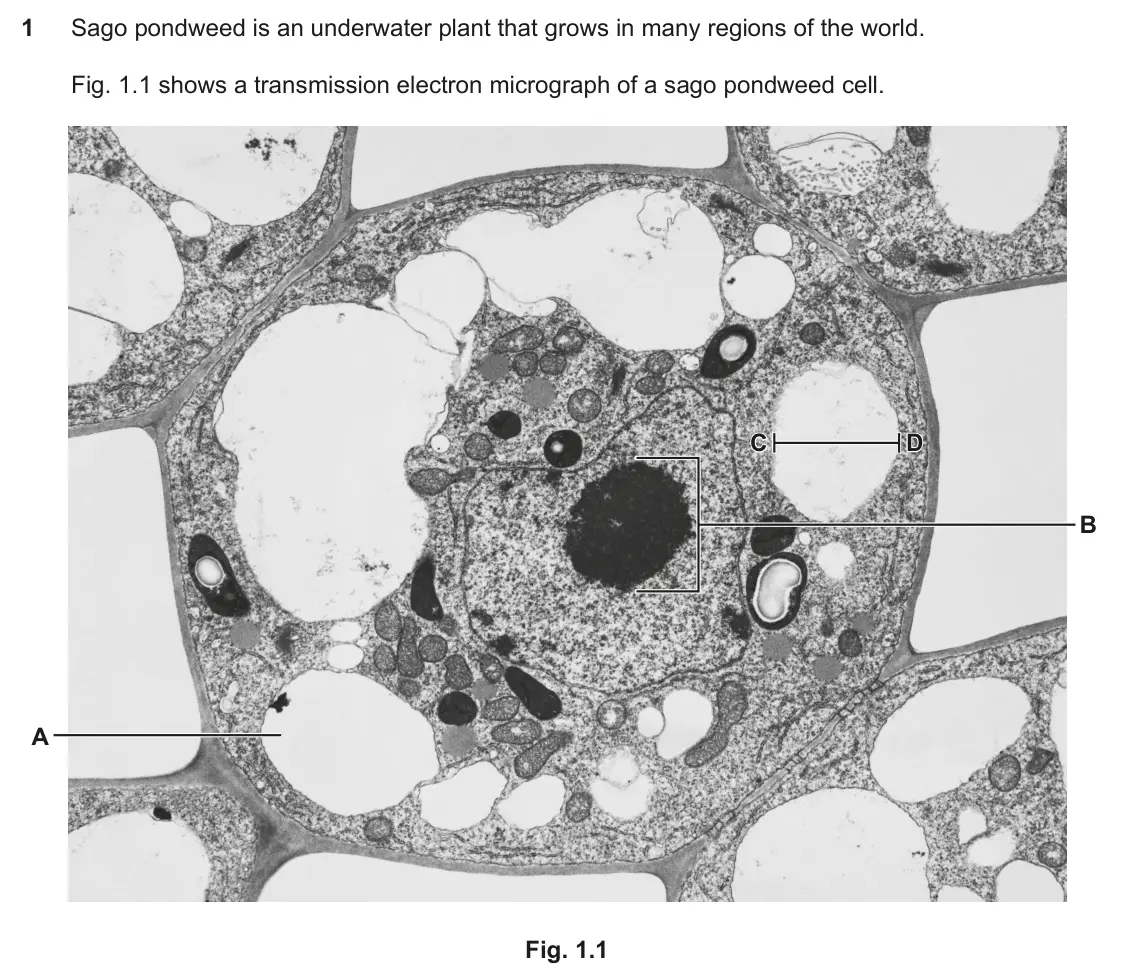
Q1(a)(iv)
The whole paper ↗Q2(c)(i)
The whole paper ↗
Q2(c)(ii)
The whole paper ↗Q16(a)(ii)
The whole paper ↗
All 22 questions on Light microscopy and slide preparation, as a list
- June 2025, Paper 2, Q16(a)(i) (2 marks): Identify two pieces of evidence that suggest that the image was produced by a light microscope rather than a scanning electron microscope. 1 2 [2]
- June 2025, Paper 2, Q16(a)(iii) (2 marks): Explain why staining is useful in microscopy. Use structures visible in the image to support your answer. [2]
- June 2025, AS Paper 1, Q26(b) (2 marks): A student is studying erythrocytes and neutrophils in a blood sample. They smear a drop of the blood sample across a microscope slide and place it under a light
- June 2024, AS Paper 2, Q5(a)(i) (2 marks): Some students collect a sample of pond water to observe under a light microscope. They pour some of the pond water onto a microscope slide. Describe how they co
- June 2024, AS Paper 2, Q5(a)(ii) (2 marks): Explain how the students would use the different objective lenses of a light microscope to focus and observe the pond water sample at high power magnification.
- June 2024, AS Paper 2, Q5(b) (2 marks): Fig. 5.1 shows a photomicrograph of human cheek cells observed by the students under a light microscope. Fig. 5.1 Magnification ×400 Fig. 5.2 shows a drawing ma
- June 2022, Paper 3, Q2(b)(iv) (1 mark): The scientist stained the lysosomes in a sample of living white blood cells. The table shows the properties of five stains, A to E. Stain Properties Suitable to
- June 2022, Paper 3, Q2(c) (2 marks): Differential staining can be used to distinguish between bacteria with thick cell walls and bacteria with thin cell walls. Four substances are used when differe
- June 2022, AS Paper 1, Q25(b)(ii) (3 marks): Name the type of microscope used to produce the photomicrograph and explain the reasons for your choice. Name of microscope Reasons for your choice [3]
- June 2022, AS Paper 2, Q2(b)(i) (1 mark): Describe how Fig. 2.4 shows the importance of differential staining for observing cells undergoing mitosis. [1]
- November 2021, AS Paper 1, Q7 (1 mark): Which of the stains, A to D, would be chosen to bind to the phosphate group of DNA to make chromosomes more visible when using a light microscope? A carbolfuchs
- November 2020, Paper 1, Q1 (1 mark): Which of the following statements, A to D, about differential staining is not true? A Differential staining can distinguish between different organelles. B Diff
- November 2020, Paper 1, Q18(b)(i) (1 mark): Explain one advantage of using a confocal microscope in this study. [1]
- November 2020, AS Paper 2, Q1(a)(i) (3 marks): Identify one incorrect label and explain your answer. Incorrect label Explanation [3]
- November 2020, AS Paper 2, Q1(a)(ii) (3 marks): State three changes, other than to the labels, to Fig. 1.2 that the student would need to make to improve the biological drawing. 1 2 3 [3]
- June 2019, Paper 3, Q1(a)(iii) (2 marks): Fig. 1.2 shows a student’s drawing of another sago pondweed cell, which was observed under a light microscope. The student used a sharp pencil but did not label
- June 2019, Paper 3, Q1(a)(iv) (2 marks): The student stained a sago pondweed sample to improve the contrast between cellular components when viewed under a microscope. The student used the following pr
- June 2019, AS Paper 2, Q2(c)(i) (3 marks): Explain why it is important to use a differential stain when examining a blood smear under the microscope. [3]
- June 2019, AS Paper 2, Q2(c)(ii) (4 marks): Using Fig. 2 on the insert, and the information provided, suggest and explain why the cytoplasm of cell C and cell D reacted differently to the stain. [4]
- June 2018, Paper 2, Q16(a)(ii) (4 marks): In the space provided below draw cell A. Label your drawing to show visible features. [4]
- June 2018, Paper 2, Q16(b)(ii) (2 marks): State the type of microscope that was used to view these images. Justify your answer. [2]
- June 2017, AS Paper 1, Q21(a)(ii) (1 mark): Suggest a stain that the student could have used to highlight the nuclei of these cells. [1]
Questions, mark schemes and examiner’s reports are © OCR, from their published past papers. Plumi isn’t affiliated with any exam board. Want whole papers? OCR A-Level Biology A past papers, or our free OCR-style practice pack.
The Plumi iPad app also offers exam-style questions with AI feedback on your handwritten working.