 plumi
Coordination and the brain: Edexcel A‑Level Biology A questions
Every Edexcel A-Level Biology A exam question on Coordination and the brain (specification points 8.7–8.9), 2016–2025, with its mark scheme and examiner’s report.
Find questions1,749
Calculations
Q1(b)(i)
The whole paper ↗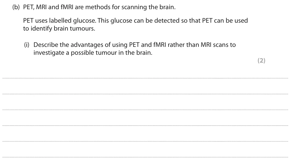
Q1(b)(ii)
The whole paper ↗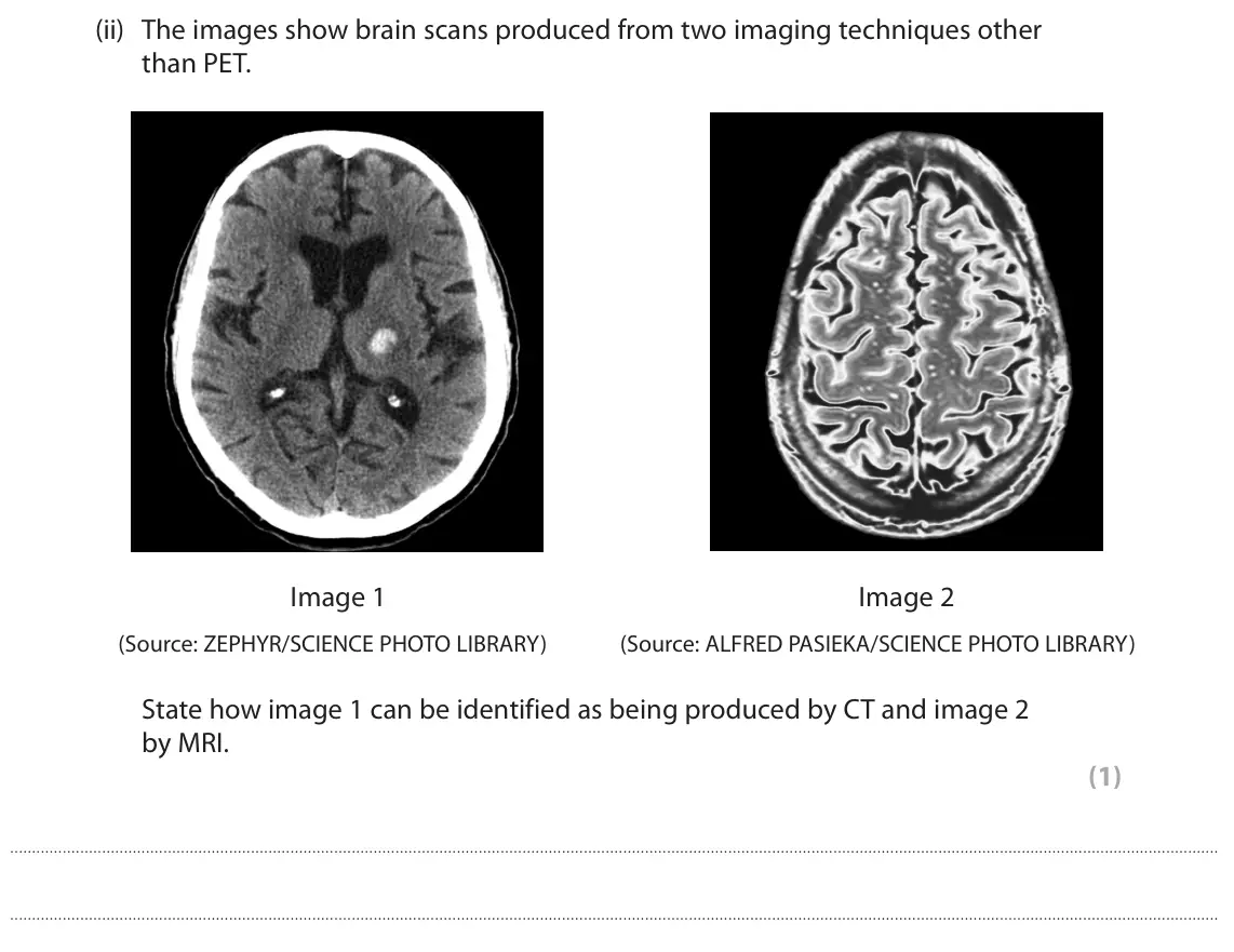
Q1(c)
The whole paper ↗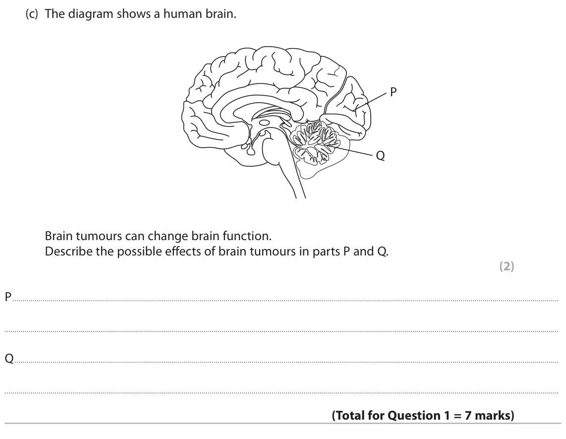
Q2(a)
The whole paper ↗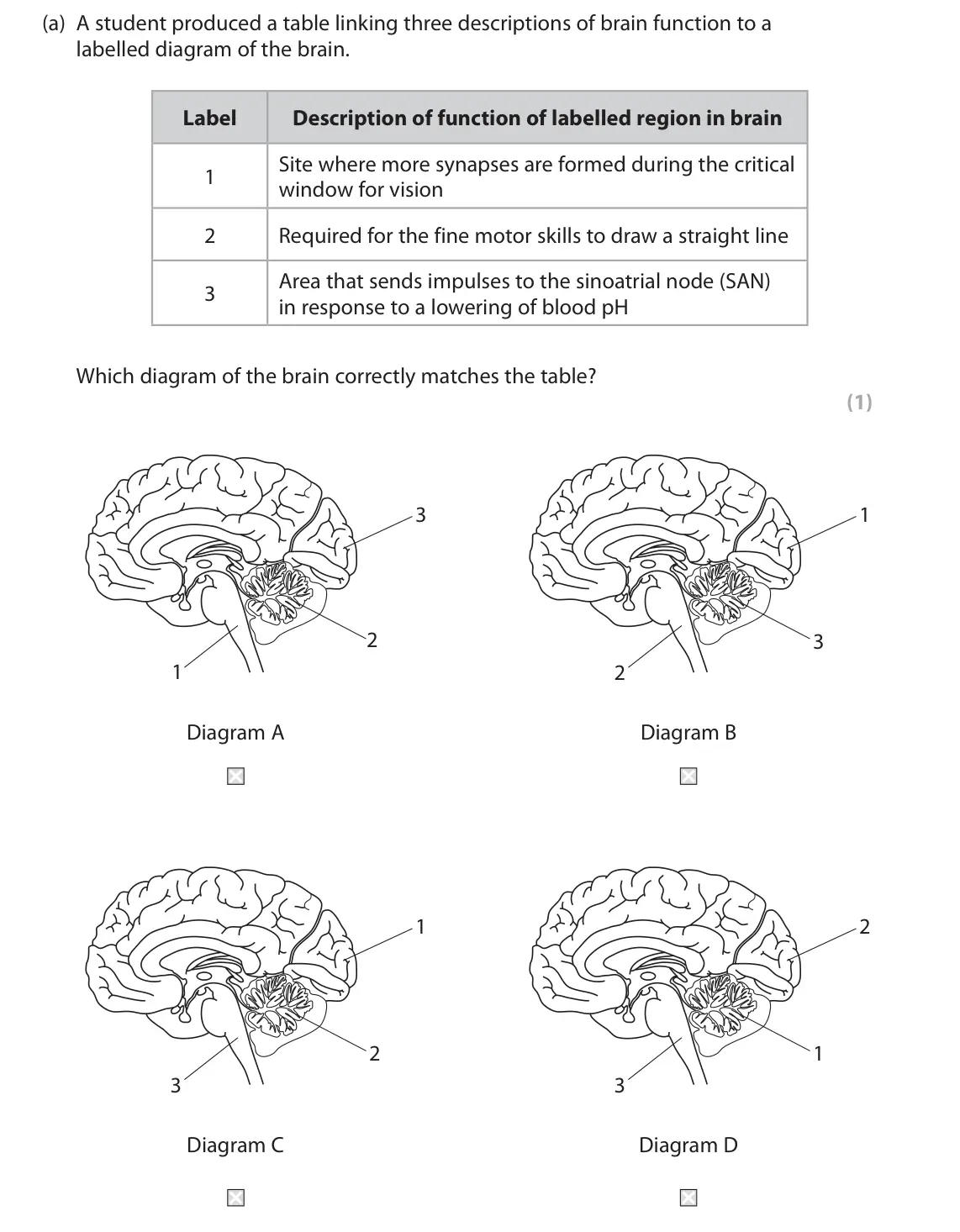
Q2(b)(i)
The whole paper ↗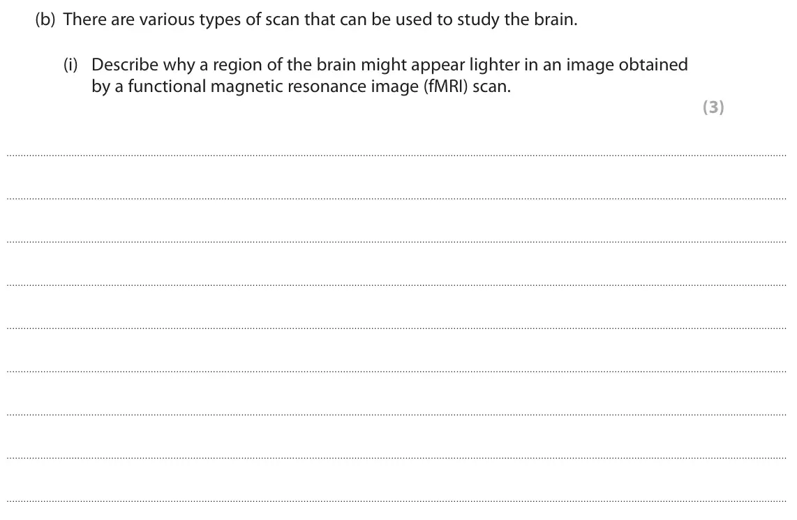
Q2(b)(ii)
The whole paper ↗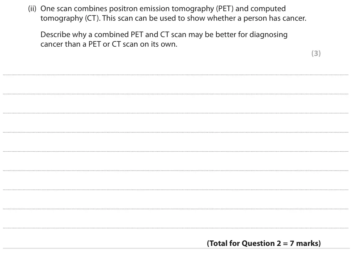
Q8(c)
The whole paper ↗
Q10(b)(ii)
The whole paper ↗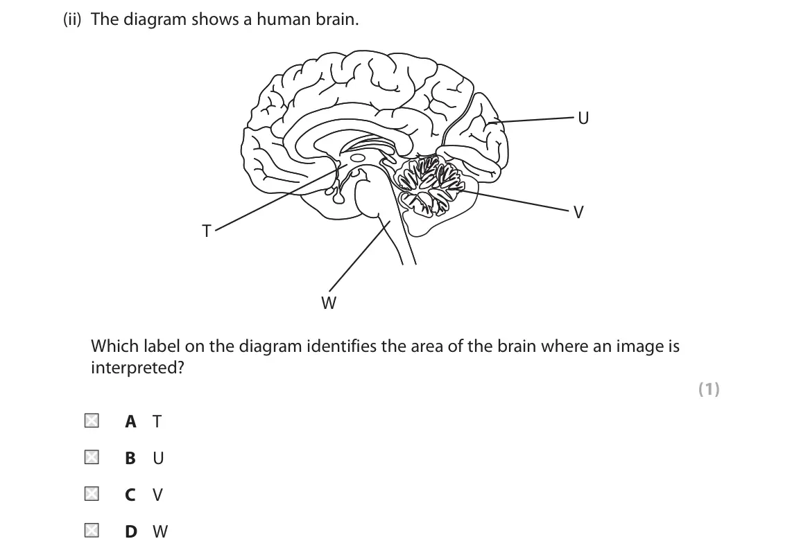
Q8(b)
The whole paper ↗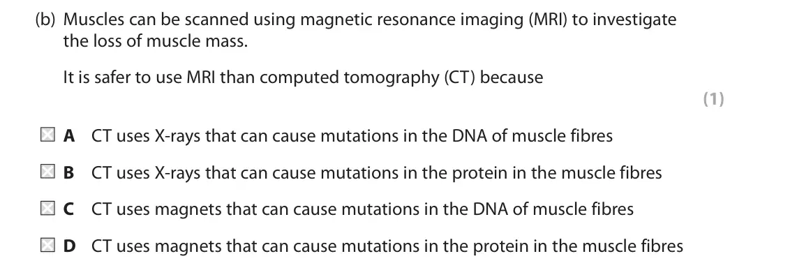
Q2(a)(i)
The whole paper ↗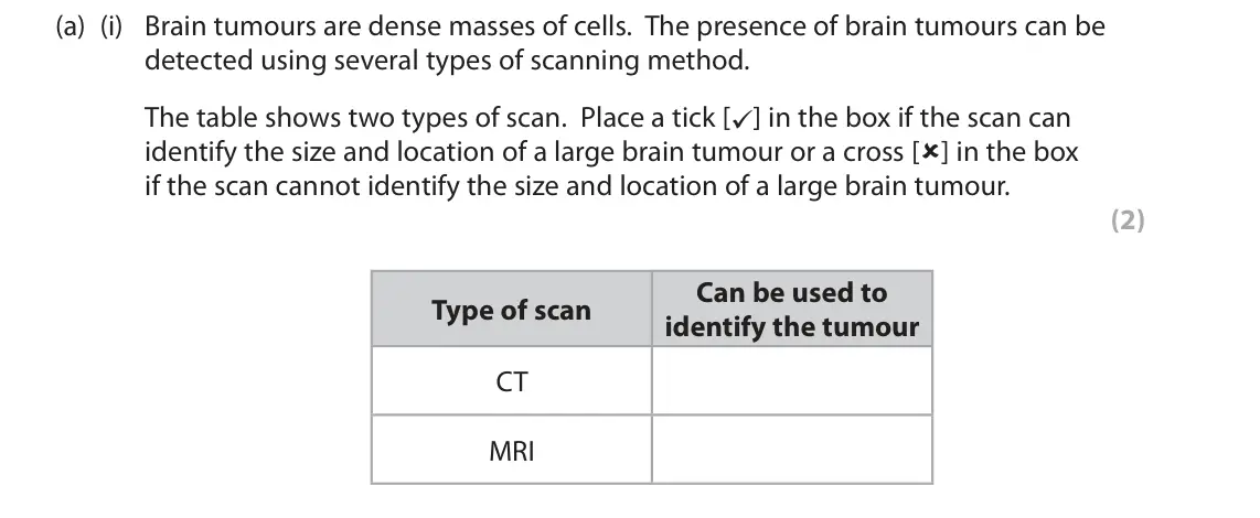
Q2(a)(ii)
The whole paper ↗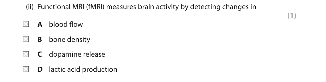
Q2(a)(iii)
The whole paper ↗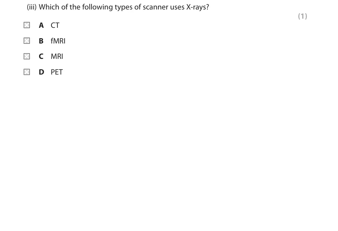
Q2(b)
The whole paper ↗
Q4(a)(ii)
The whole paper ↗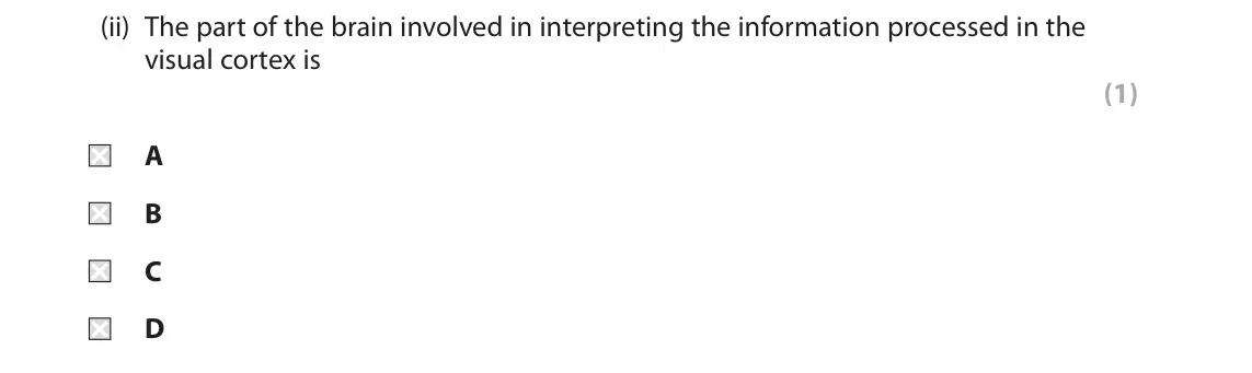
Q4(a)(iii)
The whole paper ↗
All 15 questions on Coordination and the brain, as a list
- June 2024, Paper 2, Q1(b)(i) (2 marks): Describe the advantages of using PET and fMRI rather than MRI scans to investigate a possible tumour in the brain. (2)
- June 2024, Paper 2, Q1(b)(ii) (1 mark): The images show brain scans produced from two imaging techniques other than PET. Image 1 Image 2 (Source: ZEPHYR/SCIENCE PHOTO LIBRARY) (Source: ALFRED PASIEKA/
- June 2024, Paper 2, Q1(c) (2 marks): The diagram shows a human brain. P Q Brain tumours can change brain function. Describe the possible effects of brain tumours in parts P and Q. (2) P Q
- June 2022, Paper 2, Q2(a) (1 mark): A student produced a table linking three descriptions of brain function to a labelled diagram of the brain. Label Description of function of labelled region in
- June 2022, Paper 2, Q2(b)(i) (3 marks): Describe why a region of the brain might appear lighter in an image obtained by a functional magnetic resonance image (fMRI) scan. (3)
- June 2022, Paper 2, Q2(b)(ii) (3 marks): One scan combines positron emission tomography (PET) and computed tomography (CT). This scan can be used to show whether a person has cancer. Describe why a com
- June 2022, Paper 3, Q8(c) (2 marks): Describe how tumour shrinkage could be observed (paragraph 6). (2)
- November 2021, Paper 2, Q10(b)(ii) (1 mark): The diagram shows a human brain. U V T W Which label on the diagram identifies the area of the brain where an image is interpreted? (1) A T B U C V D W
- October 2020, Paper 2, Q8(b) (1 mark): Muscles can be scanned using magnetic resonance imaging (MRI) to investigate the loss of muscle mass. It is safer to use MRI than computed tomography (CT) becau
- June 2019, Paper 2, Q2(a)(i) (2 marks): Brain tumours are dense masses of cells. The presence of brain tumours can be detected using several types of scanning method. The table shows two types of scan
- June 2019, Paper 2, Q2(a)(ii) (1 mark): Functional MRI (fMRI) measures brain activity by detecting changes in (1) A blood flow B bone density C dopamine release D lactic acid production
- June 2019, Paper 2, Q2(a)(iii) (1 mark): Which of the following types of scanner uses X-rays? (1) A CT B fMRI C MRI D PET
- June 2019, Paper 2, Q2(b) (2 marks): Describe how positron emission tomography (PET) scans can be used to investigate brain structure. (2)
- June 2018, Paper 2, Q4(a)(ii) (1 mark): The part of the brain involved in interpreting the information processed in the visual cortex is (1) A B C D
- June 2018, Paper 2, Q4(a)(iii) (3 marks): Explain how fMRI can be used to identify the part of the brain involved in interpreting information from the visual cortex. (3)
Questions, mark schemes and examiner’s reports are © Pearson Edexcel, from their published past papers. Plumi isn’t affiliated with any exam board. Want whole papers? Every Edexcel A-Level Biology A paper.
Rather write your answers by hand on iPad? Plumi marks them against the same mark schemes.