 plumi
Cell structure: AQA A‑Level Biology questions
Every AQA A-Level Biology exam question on 3.2.1 Cell structure, 2016–2025, with its mark scheme and examiner’s report.
Find questions1,383
Calculations
Q04.1
The whole paper ↗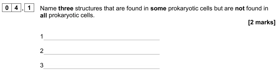
Q04.2
The whole paper ↗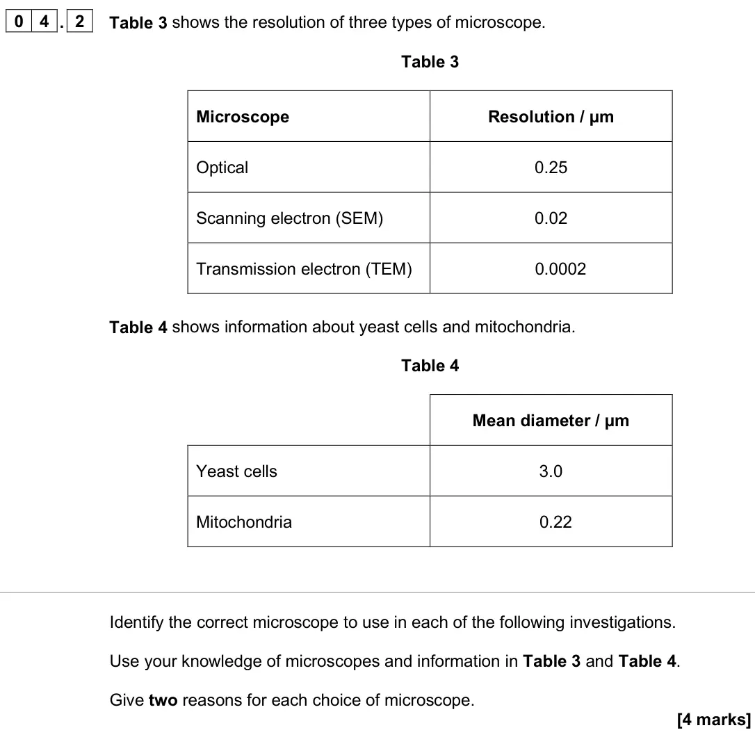
Q09.1
The whole paper ↗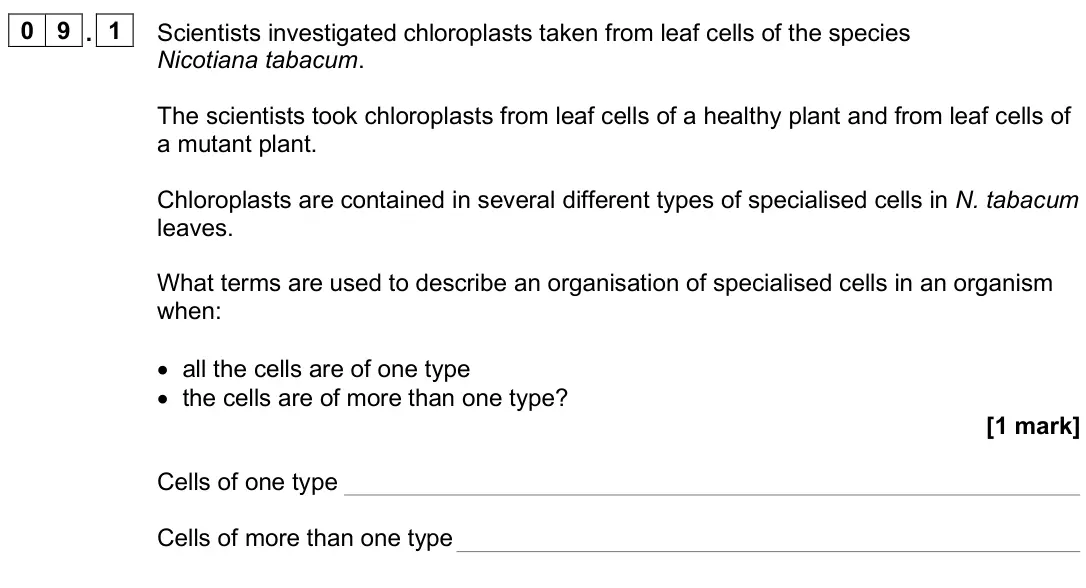
Q09.2
The whole paper ↗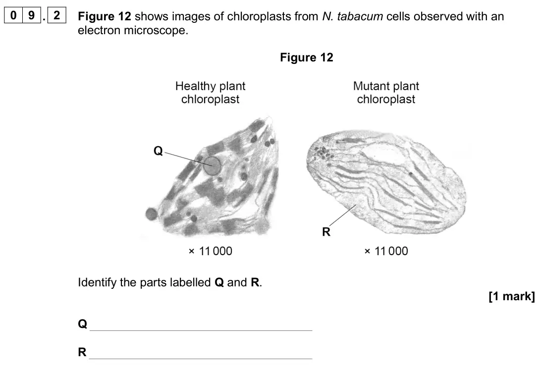
Q02.1
The whole paper ↗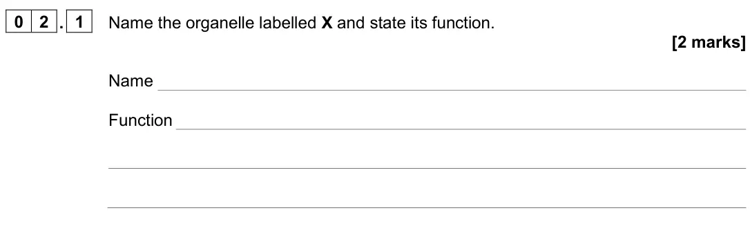
Q02.2
The whole paper ↗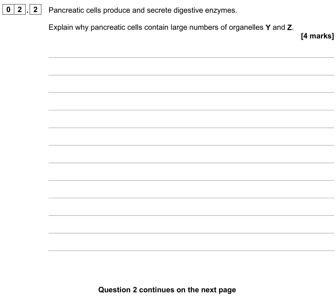
Q05.1
The whole paper ↗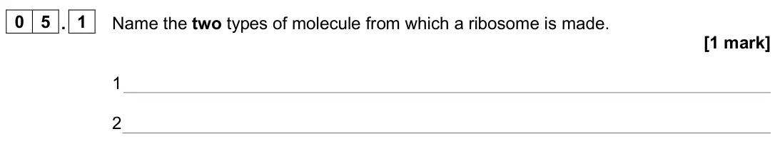
Q07.1
The whole paper ↗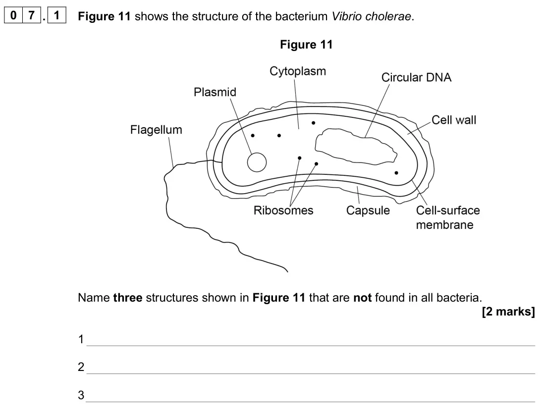
Q03.2
The whole paper ↗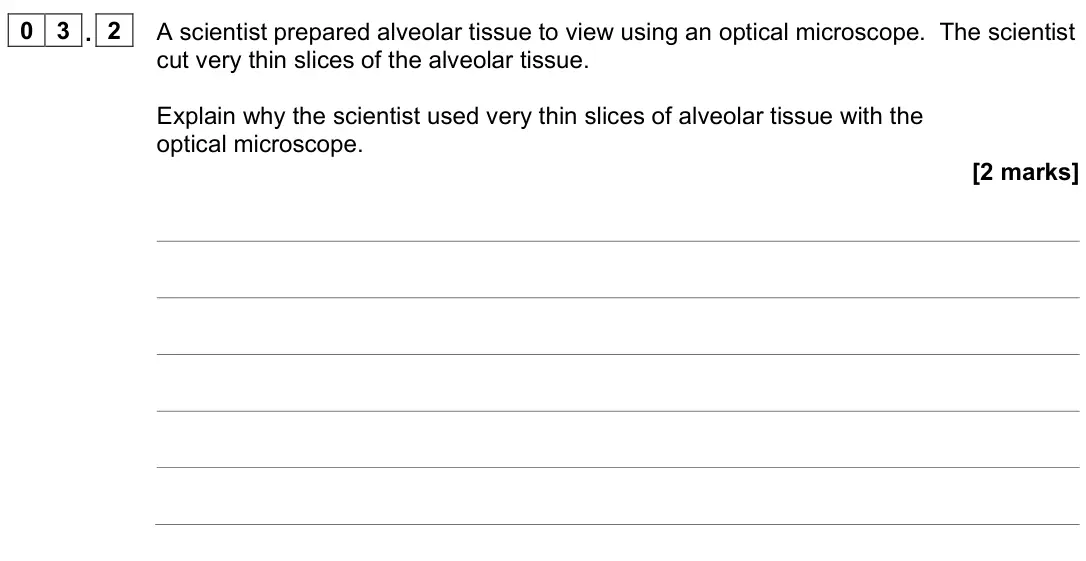
Q03.4
The whole paper ↗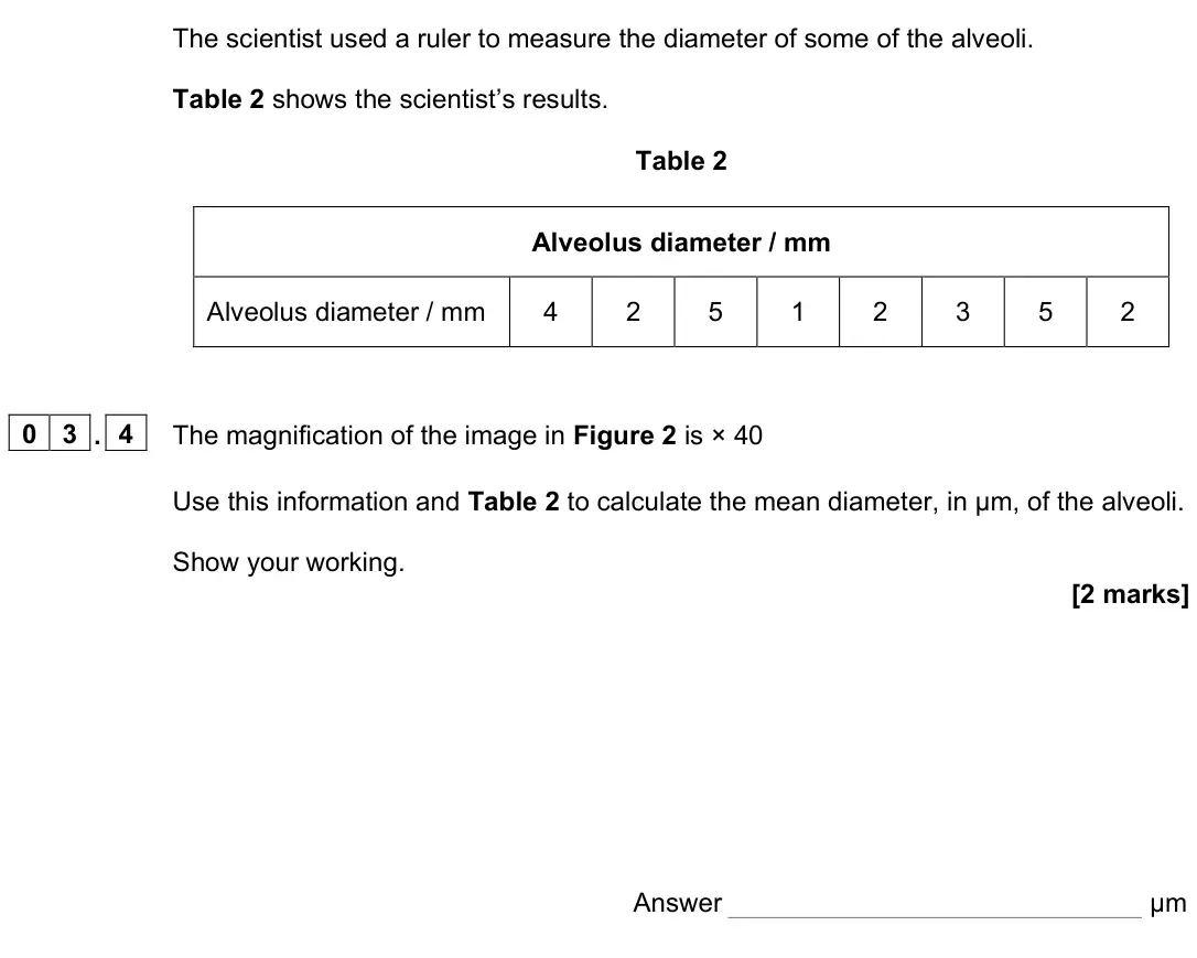
Q03.5
The whole paper ↗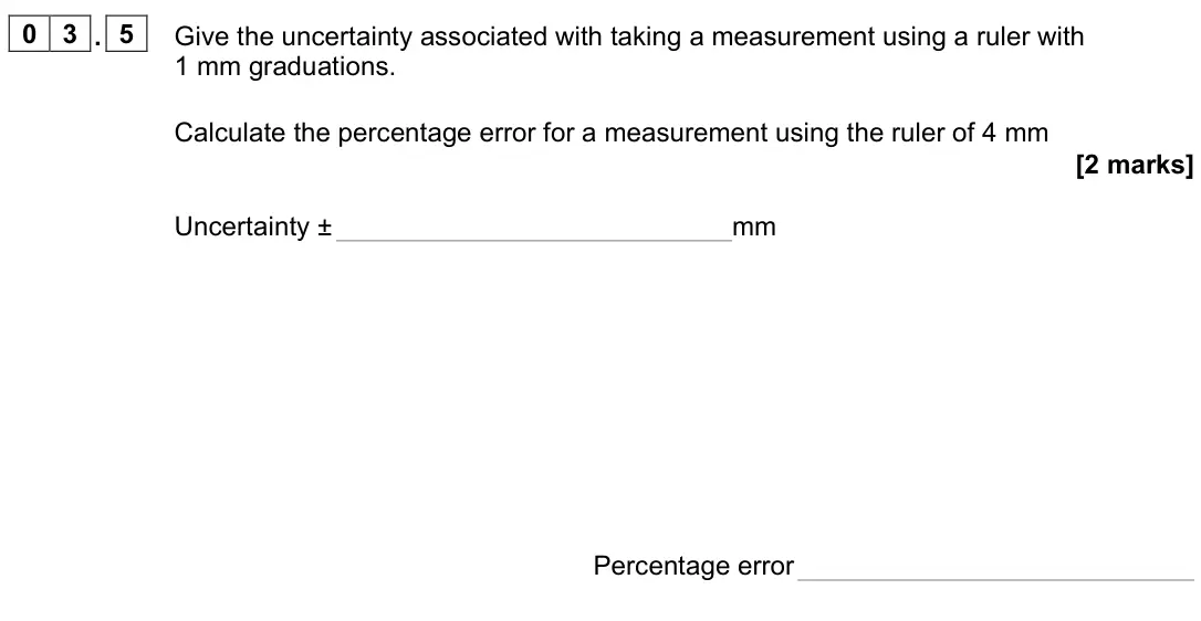
Q10.1
The whole paper ↗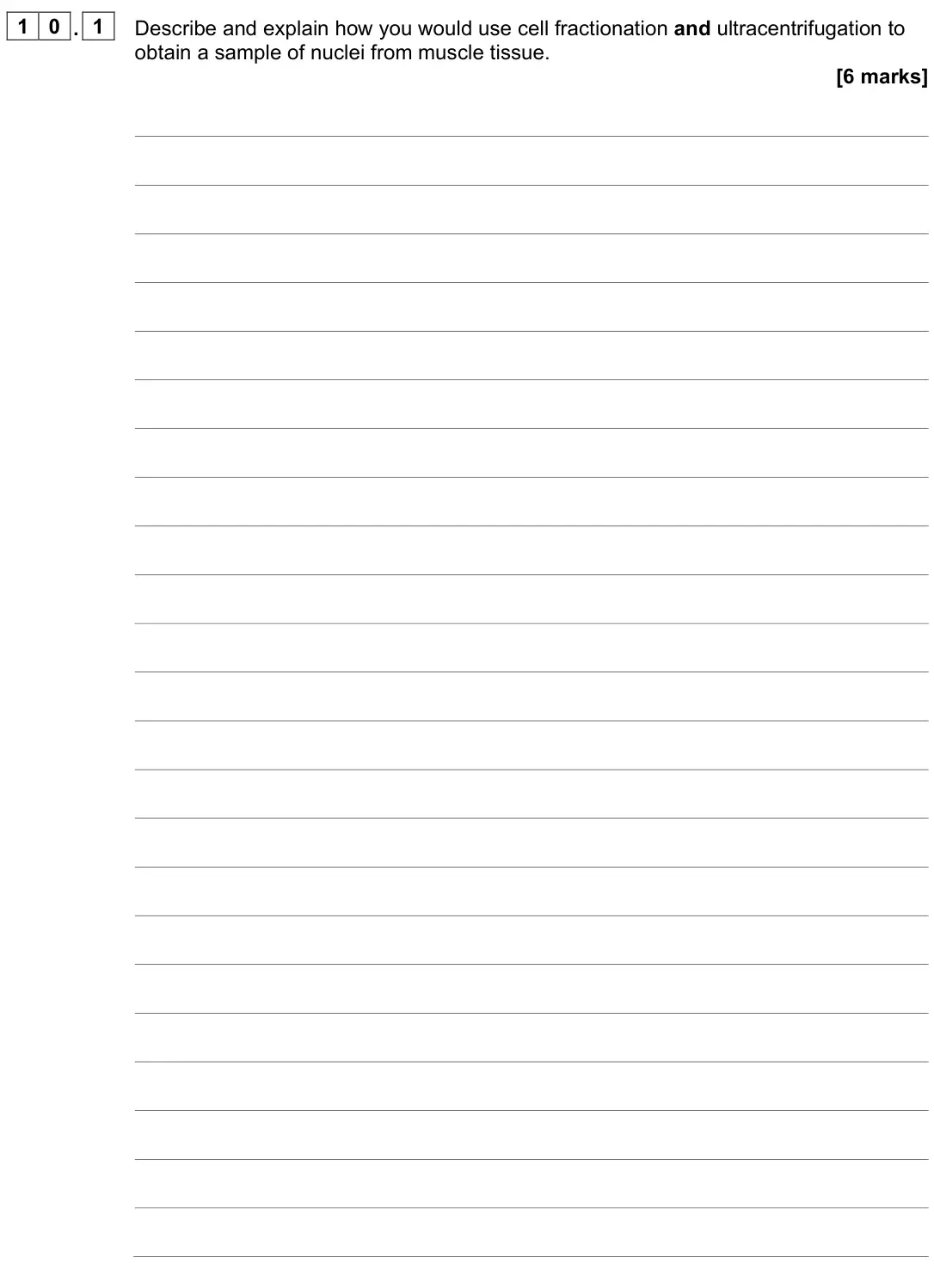
Q10.2
The whole paper ↗
Q02.1
The whole paper ↗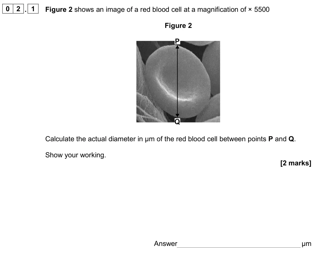
Q02.2
The whole paper ↗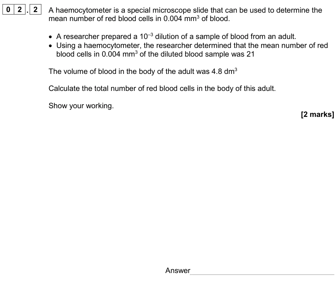
Q03.1
The whole paper ↗
Q05.4
The whole paper ↗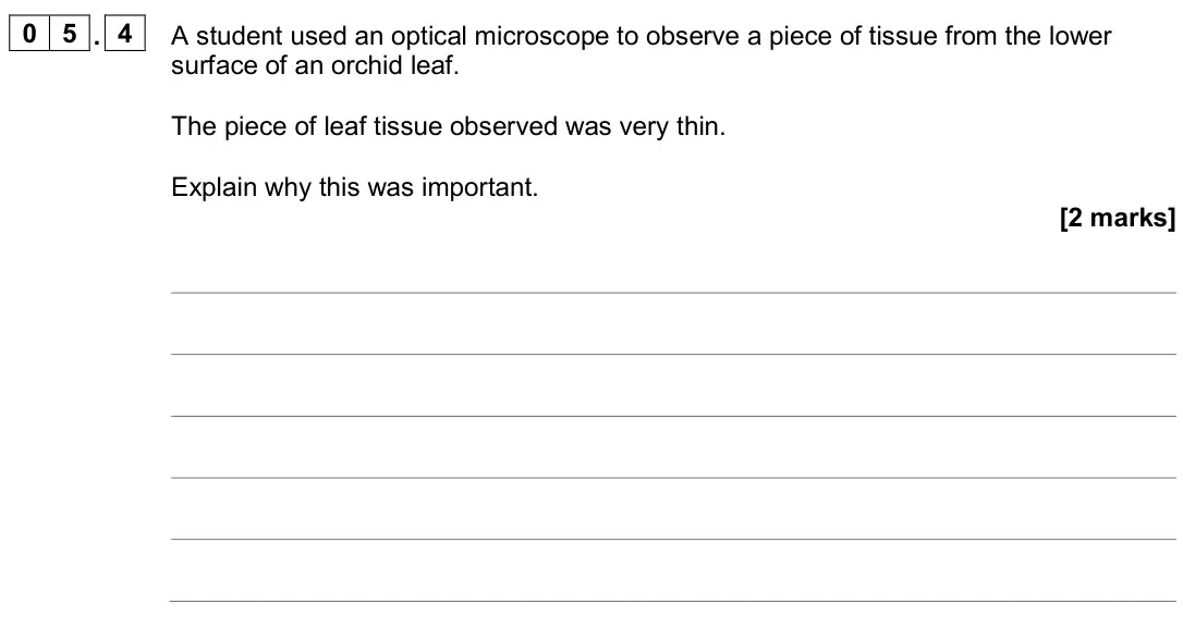
Q05.5
The whole paper ↗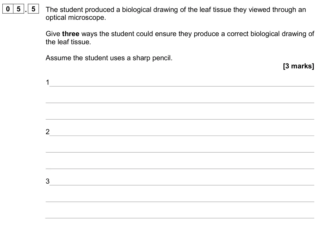
Q01.1
The whole paper ↗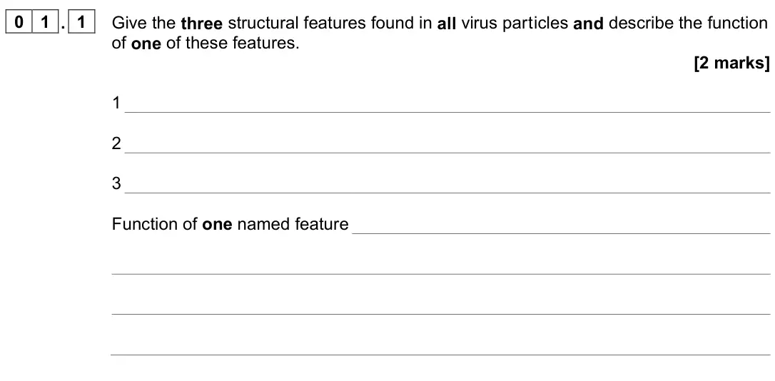
Q01.2
The whole paper ↗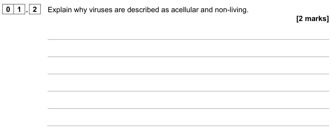
All 76 questions on Cell structure, as a list
- June 2025, Paper 1, Q04.1 (2 marks): Name three structures that are found in some prokaryotic cells but are not found in all prokaryotic cells. [2 marks] 1 2 3
- June 2025, Paper 1, Q04.2 (4 marks): Table 3 shows the resolution of three types of microscope. Table 3 Microscope Resolution / μm Optical 0.25 Scanning electron (SEM) 0.02 Transmission electron (T
- June 2025, Paper 1, Q09.1 (1 mark): Scientists investigated chloroplasts taken from leaf cells of the species Nicotiana tabacum. The scientists took chloroplasts from leaf cells of a healthy plant
- June 2025, Paper 1, Q09.2 (1 mark): Figure 12 shows images of chloroplasts from N. tabacum cells observed with an electron microscope. Figure 12 Identify the parts labelled Q and R. [1 mark] Q R
- June 2025, AS Paper 2, Q02.1 (2 marks): Name the organelle labelled X and state its function. [2 marks] Name Function
- June 2025, AS Paper 2, Q02.2 (4 marks): Pancreatic cells produce and secrete digestive enzymes. Explain why pancreatic cells contain large numbers of organelles Y and Z. [4 marks]
- June 2025, AS Paper 2, Q05.1 (1 mark): Name the two types of molecule from which a ribosome is made. [1 mark] 1 2
- June 2025, AS Paper 2, Q07.1 (2 marks): Figure 11 shows the structure of the bacterium Vibrio cholerae. Figure 11 Name three structures shown in Figure 11 that are not found in all bacteria. [2 marks]
- June 2024, Paper 1, Q03.2 (2 marks): A scientist prepared alveolar tissue to view using an optical microscope. The scientist cut very thin slices of the alveolar tissue. Explain why the scientist u
- June 2024, Paper 1, Q03.4 (2 marks): The magnification of the image in Figure 2 is × 40 Use this information and Table 2 to calculate the mean diameter, in μm, of the alveoli. Show your working. [2
- June 2024, Paper 1, Q03.5 (2 marks): Give the uncertainty associated with taking a measurement using a ruler with 1 mm graduations. Calculate the percentage error for a measurement using the ruler
- June 2024, Paper 1, Q10.1 (6 marks): Describe and explain how you would use cell fractionation and ultracentrifugation to obtain a sample of nuclei from muscle tissue. [6 marks]
- June 2024, Paper 1, Q10.2 (5 marks): Describe the role of organelles in the production and release of enzymes by animal cells. Do not include details of transcription in your answer. [5 marks]
- June 2024, AS Paper 2, Q02.1 (2 marks): Figure 2 shows an image of a red blood cell at a magnification of × 5500 Figure 2 Calculate the actual diameter in μm of the red blood cell between points P and
- June 2024, AS Paper 2, Q02.2 (2 marks): A haemocytometer is a special microscope slide that can be used to determine the mean number of red blood cells in 0.004 mm3 of blood. • A researcher prepared a
- June 2024, AS Paper 2, Q03.1 (4 marks): Describe how you would use cell fractionation and ultracentrifugation to obtain a sample of mitochondria from muscle tissue. [4 marks]
- June 2024, AS Paper 2, Q05.4 (2 marks): A student used an optical microscope to observe a piece of tissue from the lower surface of an orchid leaf. The piece of leaf tissue observed was very thin. Exp
- June 2024, AS Paper 2, Q05.5 (3 marks): The student produced a biological drawing of the leaf tissue they viewed through an optical microscope. Give three ways the student could ensure they produce a
- June 2023, Paper 1, Q01.1 (2 marks): Give the three structural features found in all virus particles and describe the function of one of these features. [2 marks] 1 2 3 Function of one named featur
- June 2023, Paper 1, Q01.2 (2 marks): Explain why viruses are described as acellular and non-living. [2 marks]
- June 2023, Paper 1, Q01.3 (1 mark): Give one reason why antibiotics are not effective against viruses. [1 mark]
- June 2023, Paper 1, Q03.1 (1 mark): The human disease, malaria, is caused by infection with a single-celled eukaryotic organism. Figure 2 shows a diagram of Plasmodium vivax, one of the species th
- June 2023, Paper 1, Q03.2 (2 marks): Describe two functions of the Golgi apparatus in a eukaryotic cell. [2 marks] 1 2
- June 2023, Paper 1, Q06.4 (2 marks): Figure 6 shows an image from an optical microscope of a single bacterial cell. Figure 6 This bacterial cell is 2.3 μm long (excluding the flagellum). Calculate
- June 2023, Paper 3, Q03.1 (1 mark): Below are four statements about the structure of prokaryotic cells. 1. No prokaryotic cell has DNA that is associated with proteins. 2. No prokaryotic cell has
- June 2023, Paper 3, Q04.1 (2 marks): Suggest how SCFR is destroyed by a lysosome. [2 marks] After a heart attack, cardiomyocytes (cardiac muscle cells) die, and become infarcted tissue. Infarcted t
- June 2023, AS Paper 1, Q02.1 (2 marks): Describe how the appearance of the area containing DNA in a TEM image of a eukaryotic cell would differ from that shown in Figure 1. [2 marks]
- June 2023, AS Paper 1, Q04.2 (2 marks): State one feature that shows the images in Figure 5 were taken with an electron microscope and not an optical microscope. Explain your answer. [2 marks]
- June 2023, AS Paper 1, Q06.1 (1 mark): Which components are found in a human papilloma virus? Tick () one box. [1 mark] Capsid and attachment protein Capsid, attachment protein and reverse transcrip
- June 2023, AS Paper 2, Q01.1 (1 mark): Which statement about the function of ribosomes is correct? Tick () one box. [1 mark] Site of transcription, catalyse the joining of amino acids by hydrolysis
- June 2023, AS Paper 2, Q01.3 (3 marks): Scientists investigated the structure of the endoplasmic reticulum. Table 1 shows some of the scientists’ results. Table 1 Type of endoplasmic reticulum Percent
- June 2023, AS Paper 2, Q10.2 (4 marks): Outline the similarities in, and the differences between, the structures of chloroplasts and mitochondria. [4 marks]
- June 2022, Paper 1, Q01.1 (4 marks): Describe the structure and function of the nucleus. [4 marks]
- June 2022, Paper 1, Q01.2 (1 mark): Name the main polymer that forms the following cell walls. [1 mark] Plant cell wall Fungal cell wall
- June 2022, Paper 1, Q03.1 (1 mark): Give two features of all prokaryotic cells that are not features of eukaryotic cells. [1 mark] 1 2
- June 2022, Paper 1, Q07.2 (2 marks): The scientists broke open the cells to produce a suspension of cell contents. Describe how the scientists would remove large organelles from this suspension of
- June 2022, Paper 1, Q07.3 (3 marks): Explain the position of the bands of ribosomes in tubes A and B in Figure 10. [3 marks] A B
- June 2022, Paper 1, Q08.1 (2 marks): To observe the fish gills with the optical microscope, the scientists used two different stains. The first stain binds to DNA; the second stain binds to the red
- June 2022, AS Paper 1, Q02.1 (2 marks): Give the two types of molecule from which a ribosome is made. [2 marks] 1 2
- June 2022, AS Paper 1, Q03.1 (1 mark): Figure 2 is an image of a bacterium obtained using a scanning electron microscope. Figure 2 Name the structure labelled X. [1 mark]
- June 2022, AS Paper 1, Q03.2 (2 marks): Figure 2 is different from an image of this bacterium obtained using a transmission electron microscope. Describe and explain one difference between these image
- June 2022, AS Paper 1, Q03.3 (1 mark): The resolution of an image obtained using an electron microscope is higher than the resolution of an image obtained using an optical microscope. Explain why. [1
- June 2022, AS Paper 1, Q03.4 (2 marks): A student determined the size of a cell structure from a photograph obtained using a microscope. He used a ruler and a calculator and gave the answer in μm Desc
- June 2022, AS Paper 1, Q03.5 (2 marks): Name two structures found in all bacteria that are not found in plant cells. [2 marks] 1 2
- June 2022, AS Paper 1, Q03.6 (2 marks): Name two features of HIV particles that are not found in bacteria. Do not include attachment protein in your answer. [2 marks] 1 2
- June 2022, AS Paper 2, Q07.1 (3 marks): In the box below: • produce a scientific drawing of the arrangement of tissues shown in Figure 8 • label one of the tissues in your drawing Do not draw individu
- November 2021, Paper 1, Q05.1 (4 marks): Describe how a sample of chloroplasts could be isolated from leaves. [4 marks]
- November 2021, Paper 1, Q05.4 (3 marks): The scientists also observed the chloroplasts from the samples of leaves using an electron microscope. Figure 6 shows a chloroplast from a control plant (image
- November 2021, Paper 1, Q10.3 (5 marks): Contrast the structure of the two cells visible in the electron micrographs shown in Figure 14. [5 marks]
- November 2021, Paper 2, Q07.1 (2 marks): HBV infects a liver cell. The liver cell is 25 µm in diameter. Use Figure 7 to calculate how many times larger in diameter this cell is than HBV. You should use
- November 2021, AS Paper 1, Q02.1 (2 marks): Name structures A and B. [2 marks] A B
- November 2021, AS Paper 1, Q03.1 (2 marks): Explain why it is not possible to determine the identity of the structures labelled X using an optical microscope. [2 marks]
- November 2021, AS Paper 1, Q03.2 (3 marks): U. marinum cells ingest bacteria and digest them in the cytoplasm. Describe the role of one named organelle in digesting these bacteria. [3 marks]
- November 2021, AS Paper 1, Q03.3 (2 marks): Calculate the actual length of the cell shown between Y and Z in Figure 3. The magnification of the image is × 900 Give your answer in μm and to 2 significant f
- November 2021, AS Paper 1, Q09.1 (1 mark): Kidney cells produce a glycoprotein called erythropoietin (EPO) (line 1). Identify two organelles in kidney cells that enable the production of EPO. [1 mark] 1
- November 2021, AS Paper 2, Q04.1 (2 marks): Give two structures found in all prokaryotic cells and in all eukaryotic cells. [2 marks] 1 2
- November 2021, AS Paper 2, Q04.3 (2 marks): What can you conclude from Figure 6 about a structural difference between the plasmids and the circular DNA? Explain your answer. [2 marks]
- November 2020, AS Paper 1, Q04.1 (4 marks): Eukaryotic cells produce and release proteins. Outline the role of organelles in the production, transport and release of proteins from eukaryotic cells. Do not
- November 2020, AS Paper 1, Q04.2 (1 mark): Suggest why a nucleus is not visible in Figure 3. [1 mark]
- November 2020, AS Paper 1, Q04.3 (1 mark): Name the organelles labelled S and T in Figure 3. [1 mark] Organelle S Organelle T
- November 2020, AS Paper 1, Q04.4 (1 mark): Give one advantage of viewing a biological specimen using a transmission electron microscope compared with using a scanning electron microscope. [1 mark]
- November 2020, AS Paper 1, Q04.5 (2 marks): The cells in Figure 4 are part of a continuous layer of cells forming the upper surface of a leaf. The shaded area of cell U is 150 µm2 The total area of the up
- November 2020, AS Paper 1, Q05.3 (2 marks): Two solutions often used to stain tissues are haematoxylin solution and iodine solution. • Haematoxylin solution stains DNA a blue colour. • Iodine solution sta
- June 2017, Paper 3, Q02.1 (1 mark): Using Figure 2 and your own knowledge, put a tick () in the box next to the only correct statement about the structures of the bacteriophage and the bacterium.
- June 2017, Paper 3, Q02.2 (2 marks): Using the scales in Figure 2, calculate how many times longer the bacterium is than the bacteriophage. Use the distance between the points labelled A and B on e
- June 2017, Paper 3, Q02.3 (1 mark): Scientists investigated the use of bacteriophages to treat lung infections caused by bacteria. They infected the lungs of mice with a pathogenic species of bact
- June 2017, Paper 3, Q02.4 (2 marks): With some samples, the scientists decided they needed to carry out a series of dilutions of the sample before counting the bacteria. Use evidence from Figure 3
- June 2017, Paper 3, Q02.5 (3 marks): Using only Figure 3, what can you conclude from these data about the effectiveness of the bacteriophage in treating this lung infection in mice? Do not consider
- June 2017, AS Paper 1, Q07.1 (3 marks): Describe how you would use cell fractionation techniques to obtain a sample of chloroplasts from leaf tissue. Do not include in your answer information about an
- June 2017, AS Paper 1, Q07.2 (3 marks): Table 3 shows features of a mitochondrion and a chloroplast. Complete Table 3 with ticks where a feature is present. [3 marks] Table 3 Feature Mitochondrion Chl
- June 2017, AS Paper 1, Q07.3 (1 mark): Give the function of a mitochondrion. [1 mark]
- June 2017, AS Paper 1, Q07.4 (2 marks): Scientists investigated the effect of an exercise programme on the number and size of mitochondria in skeletal muscle. They took samples of muscle from a large
- Specimen, Paper 3, Q02.1 (4 marks): [ ] This source has been removed due to third-party copyright restrictions.
- Specimen, Paper 3, Q02.2 (2 marks): Name of W Function of W Name of Z Function of Z
- Specimen, Paper 3, Q02.3 (2 marks): A transmission electron microscope was used to produce the image in Figure 1. Explain why. [2 marks] 0 2 . 3
- Specimen, Paper 3, Q02.4 (1 mark): Answer =
Questions, mark schemes and examiner’s reports are © AQA, from their published past papers. Plumi isn’t affiliated with any exam board. Want whole papers? Every AQA A-Level Biology paper.
Rather write your answers by hand on iPad? Plumi marks them against the same mark schemes.