 plumi
Cells and microscopy: OCR A‑Level Biology B questions
Every OCR A-Level Biology B exam question on 2.1.1 Cells and microscopy, 2016–2025, with its mark scheme and examiner’s report.
Find questions1,889
Calculations


Q33(b)(ii)
The whole paper ↗
Q34(a)(ii)
The whole paper ↗
Q1(a)(ii)
The whole paper ↗
Q3(a)
The whole paper ↗
Q3(b)
The whole paper ↗


Q24(b)(ii)
The whole paper ↗
Q25(a)(i)
The whole paper ↗
Q25(a)(ii)
The whole paper ↗
Q25(a)(iii)
The whole paper ↗
Q25(b)(i)
The whole paper ↗
Q25(b)(ii)
The whole paper ↗
Q25(b)(iii)
The whole paper ↗
Q25(c)
The whole paper ↗


All 139 questions on Cells and microscopy, as a list
- June 2025, Paper 1, Q1 (1 mark): Which type of blood cell is shown in the annotated drawing? Non-granulated cytoplasm Kidney-bean shaped nucleus A Erythrocyte B Lymphocyte C Monocyte D Neutroph
- June 2025, Paper 1, Q2 (1 mark): Which technique uses fluorescent markers to tag cells so they can be counted and identified when passed through a laser beam? A Differential staining B Flow cyt
- June 2025, Paper 1, Q33(b)(ii) (4 marks): The diagram below shows a type of cell that was extracted from the developing root nodule labelled in the photomicrograph. Cell wall Plasma membrane Cytoplasm D
- June 2025, Paper 1, Q34(a)(ii) (2 marks): The same myofibril has been labelled G on both micrographs. Calculate the actual diameter of myofibril G before spaceflight. Diameter = μm [2]
- June 2025, Paper 3, Q1(a)(ii) (2 marks): The diameter of one region of the root is represented by the line C in the image. The magnification used to produce the image was × 20. Calculate the actual dia
- June 2025, Paper 3, Q3(a) (2 marks): State two ways in which the student could improve their drawing. 1 2 [2]
- June 2025, Paper 3, Q3(b) (3 marks): The student concludes that ‘although it does not seem to have mitochondria, P. myojinensis is more similar to a eukaryote than a prokaryote’. Discuss the eviden
- June 2025, AS Paper 1, Q3 (1 mark): The transmission electron micrograph shows the ultrastructure of a mammalian cell. P Which option describes the function of the structure labelled P? A It produ
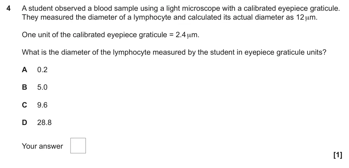
- June 2025, AS Paper 1, Q4 (1 mark): A student observed a blood sample using a light microscope with a calibrated eyepiece graticule. They measured the diameter of a lymphocyte and calculated its a
- June 2025, AS Paper 1, Q13 (1 mark): Which molecule is found in the cell wall of a bacterium? A Cellulose B Lignin C Peptidoglycan D Phospholipid Your answer [1]
- June 2025, AS Paper 1, Q24(b)(ii) (1 mark): Suggest the mechanism of transport across cell surface membranes that involves dynamin. [1] 20 In a recent study into the role of dynamin in the infection of ho
- June 2025, AS Paper 1, Q25(a)(i) (1 mark): Suggest one reason why steps 1 and 2 are included in the method. [1]
- June 2025, AS Paper 1, Q25(a)(ii) (1 mark): Suggest one reason why it is necessary to mix the sample again thoroughly in step 4. [1]
- June 2025, AS Paper 1, Q25(a)(iii) (3 marks): Outline further steps in the method that would enable the students to estimate the number of individual algae present in the sample. [3]
- June 2025, AS Paper 1, Q25(b)(i) (1 mark): Using the image, explain a disadvantage of using a haemocytometer to estimate the number of algal cells in a pond water sample. [1]
- June 2025, AS Paper 1, Q25(b)(ii) (2 marks): The diameter of individual P of the algal species, Pandorina morum, is shown on the image. Calculate the actual diameter of this individual. Diameter = μm [2]
- June 2025, AS Paper 1, Q25(b)(iii) (3 marks): Wet biomass (biovolume) of P. morum in the sample can be calculated using the equation: Biovolume = number of individuals per cm3 × mean volume of individuals T
- June 2025, AS Paper 1, Q25(c) (2 marks): The diagram shows the ultrastructure of an algal cell from the Euglena species. R S U T Give the letters identifying one organelle that would be present and one
- June 2025, AS Paper 2, Q1(b) (2 marks): The image below shows a section through the wall of an artery. X Y ×500 Calculate the actual width of the artery wall between points X and Y. Give your answer t
- June 2025, AS Paper 2, Q6(a)(i) (2 marks): State the type of microscope used to produce this image. Give a reason for your answer. Type Reason [2]
- June 2024, Paper 1, Q2 (1 mark): Which of the options identifies the leucocyte in the electron micrograph? A Lymphocyte B Macrophage C Monocyte D Neutrophil Your answer [1]
- June 2024, Paper 1, Q3 (1 mark): What is the role of the organelle labelled L? A Aerobic respiration B Lipid synthesis C Protein synthesis D Transport Your answer [1]
- June 2024, Paper 1, Q34(a)(ii) (2 marks): Calculate the actual length of Q between points X and Y. Actual length = μ m [2]
- June 2024, Paper 3, Q3(c) (1 mark): Hypothyroidism can affect the concentration of red blood cells in a person’s blood. State a method that could be used to measure the concentration of red blood
- June 2024, AS Paper 1, Q21(c)(i) (2 marks): Suggest a type of microscope used to produce the photomicrograph in Fig. 21.2 and give a reason for your choice. Type of microscope Reason [2]
- June 2024, AS Paper 1, Q21(c)(ii) (2 marks): Calculate the actual length of the 1 cm scale bar shown on Fig. 21.2 and give appropriate units. Actual length = Units = [2]
- June 2024, AS Paper 1, Q23(a)(i) (1 mark): Suggest why a buffer was used in this investigation. [1]
- June 2024, AS Paper 1, Q23(a)(ii) (1 mark): Suggest one reason why the scientists waited 200 s before adding the chloroplast solution. [1]
- June 2024, AS Paper 1, Q23(b)(i) (3 marks): Use the grid to plot an appropriate graph for these data. [3]
- June 2024, AS Paper 1, Q23(b)(ii) (2 marks): Use your graph to calculate the rate of uptake of CO2 at 275 s. Give your answer in standard form. Rate of uptake of CO2 = μmol dm–3 s–1 [2]
- June 2024, AS Paper 1, Q23(b)(iii) (3 marks): Use your graph to suggest a mechanism of transport for CO2 across the membrane and comment on the permeability of chloroplast membranes to CO2. [3]
- June 2024, AS Paper 2, Q1(a)(iii) (2 marks): Calculate the actual length of the structure between X–Y. Actual length = μm [2]
- June 2024, AS Paper 2, Q2(b)(i) (2 marks): Using your knowledge of transport of molecules across membranes, explain the shape of the curve at region A and at region B. Region A Region B [2]
- June 2024, AS Paper 2, Q2(b)(ii) (1 mark): Cells in the salivary glands require oxygen for aerobic respiration. Draw a line on the graph to show the rate of uptake of oxygen by the cells of the salivary
- June 2024, AS Paper 2, Q2(c) (6 marks): Amino acids have the same general structure with four different chemical groups attached to a central carbon atom. The structure of the amino acid valine is sho
- June 2024, AS Paper 2, Q5(d)(ii) (2 marks): Cytotoxin has both polar and non-polar regions. Suggest how cytotoxin binds to the membrane and causes damage to the blood cells. [2]
- June 2023, Paper 1, Q2 (1 mark): The diagram shows substance X being transported across a cell membrane. Substance X 2 1 Which transport mechanism is being used to transport substance X? A Acti
- June 2023, Paper 1, Q36(a)(i) (2 marks): Outline a correct method for counting cells using a haemocytometer. [2]
- June 2023, Paper 1, Q36(a)(ii) (2 marks): Explain how the class results would be affected by including the results obtained by the student who counted all the cells in their field of view. [2]
- June 2023, Paper 1, Q36(b) (3 marks): Other methods can be used to count cells. Complete the sentences about a method to count cells using the most appropriate word(s). is an electronic method of co
- June 2023, Paper 2, Q3(a)(i) (1 mark): State the letter on Fig. 3.1 that identifies the component of the membrane that binds to LPS. [1]
- June 2023, Paper 2, Q3(a)(ii) (2 marks): Explain why the membrane is described as a fluid mosaic. [2]
- June 2023, Paper 2, Q7(c) (4 marks): Lung surfactant is a mixture of phospholipids and proteins that helps the alveoli stretch and expand during breathing in and prevents them collapsing when breat
- June 2023, Paper 3, Q1(a)(ii) (2 marks): The magnification used to produce the TEM in Fig. 1.1 was × 20 000. Calculate the actual diameter of the structure labelled D in Fig. 1.1. Give your answer in s
- June 2023, Paper 3, Q4(d)(ii) (6 marks): The domestic rabbit, Oryctolagus cuniculus domesticus, is a subspecies of European rabbit. It is a popular pet and has been selectively bred for fur colour and
- June 2022, Paper 1, Q11 (1 mark): Flow cytometry can be used to determine the concentration of bacterial cells in a sample by comparing fluorescently-stained bacterial cells with a known concent
- June 2022, Paper 1, Q31(c)(ii) (2 marks): Daptomycin has a bactericidal effect on gram positive bacteria by causing cytosol to leak out of bacterial cells. Using the information in Fig. 31.2, suggest ho
- June 2022, Paper 1, Q35(a)(i) (1 mark): Name the type of leucocyte shown. [1]
- June 2022, Paper 1, Q35(a)(ii) (2 marks): Make an annotated drawing of the leucocyte. [2] 35 The eyepiece graticule used was calibrated as shown in the diagram. 100 μm 0 10 20 30 40 50 60 70 80 90100 Ey
- June 2022, Paper 1, Q35(a)(iii) (1 mark): Name the apparatus used to calibrate the eyepiece graticule. [1]
- June 2022, Paper 1, Q35(a)(iv) (2 marks): Calculate the diameter of the leucocyte. Give your answer to 2 significant figures. Diameter = µm [2]
- June 2022, Paper 3, Q1(a)(iii) (2 marks): The diameter of one region of pancreatic tissue is represented by the line Z. Calculate the actual diameter of the region labelled Z. Give your answer in µm and
- June 2022, Paper 3, Q1(a)(iv) (2 marks): A stage micrometer can be used when measuring the linear dimensions of cells, such as those of the pancreatic tissue, viewed under a light microscope. Outline h
- June 2022, AS Paper 1, Q25(a) (2 marks): Identify the blood cells in Fig. 25 and complete the table below. Name Letter Erythrocyte Monocyte Lymphocyte [2]
- June 2022, AS Paper 1, Q25(b)(i) (1 mark): Give one reason why it was important to make a correct smear (film) of the blood sample. [1]
- June 2022, AS Paper 1, Q25(b)(ii) (1 mark): Give one reason why Leishman’s stain was used. [1]
- June 2022, AS Paper 1, Q25(b)(iii) (1 mark): Suggest why macrophages were not present in this blood sample. [1]
- June 2022, AS Paper 2, Q6(a)(i) (1 mark): Draw a box on Fig. 6.1 around the region of the phospholipid that is glycerol. [1]
- June 2022, AS Paper 2, Q6(a)(ii) (1 mark): Label A in Fig. 6.1 shows a double bond between carbons. Describe the effect this double bond between carbons has on the properties of this molecule. [1]
- June 2022, AS Paper 2, Q6(b)(i) (2 marks): Explain why the cell surface membrane of an intestinal epithelial cell contains cholesterol but the mitochondrial membrane does not. [2]
- June 2022, AS Paper 2, Q6(b)(ii) (1 mark): Counting the number of each type of lipid molecule would be too difficult to do accurately. Suggest one other reason why calculating the percentage mass of the
- June 2022, AS Paper 2, Q6(c) (4 marks): The rate of active transport of a molecule across a membrane is dependent on a number of different factors. State and explain two factors that can increase the
- November 2021, Paper 1, Q8 (1 mark): Milk fat contains many different types of triglycerides. The molecules that make up these triglycerides can affect the flavour and nutritional properties of mil
- November 2021, Paper 1, Q14 (1 mark): An investigation was carried out into the effect of concentration of a substance on its rate of uptake into cells. The cells were placed in solutions of the sub
- November 2021, Paper 1, Q15 (1 mark): Which of the eukaryotic cell membranes, A to D, is used to isolate enzymes that could cause cellular damage? A lysosome membrane B mitochondrial membrane C nucl
- November 2021, Paper 1, Q32(a) (2 marks): The tissues of the eye shown in the photomicrograph were stained using eosin, which binds to cytoplasmic proteins staining them pink, and haematoxylin, which st
- November 2021, Paper 1, Q32(c)(i) (2 marks): Find the mean of the data and use it to calculate the actual width of the retinal layer. Width = μm [2]
- November 2021, Paper 1, Q32(c)(ii) (2 marks): Calculate the uncertainty in the mean value for this data. Use the equation: R 2√N R = difference between maximum and minimum values N = number of measurements
- November 2021, Paper 3, Q3(b) (2 marks): Fig. 3.2 is a photomicrograph of the lower surface of a leaf showing stomata as viewed using a light microscope. P ×1200 Fig. 3.2 Calculate the actual length of
- November 2021, AS Paper 1, Q1 (1 mark): A student used a haemocytometer to count the number of yeast cells in a diluted sample. The North-West counting rule was applied by the student when counting th
- November 2021, AS Paper 1, Q18 (1 mark): Researchers used radioactively labelled amino acids to track the movement of proteins through a The graph below shows the percentage of radioactively labelled p
- November 2021, AS Paper 2, Q1(a)(ii) (2 marks): Calculate the magnification of the image in Fig. 1.1. Magnification = [2]
- November 2021, AS Paper 2, Q1(d) (6 marks): Fig. 1.1 is a photomicrograph showing some of the cells that line the bronchus. X 4 μm Fig. 1.1 (d)* Eosinophils are a type of white blood cell involved in prod
- November 2021, AS Paper 2, Q6(a) (2 marks): Describe two structural differences between an erythrocyte and a prokaryotic cell such as M. tuberculosis. Difference 1 Difference 2 [2] Question 6(b) begins on
- November 2021, AS Paper 2, Q6(b) (3 marks): Flow cytometry is a technique used to detect and measure the physical characteristics of blood cells and platelets. The graph shows the analysis of a blood samp
- November 2020, Paper 1, Q2 (1 mark): Which of the molecules, A to D, reduces the permeability of the plasma membrane to most substances? A carbohydrate B cholesterol C glycoprotein D protein Your a
- November 2020, Paper 1, Q3 (1 mark): To which of the substances, A to D, is the plasma membrane of an axon least permeable? A ATP B glucose K+ C Na+ D Your answer [1]
- November 2020, Paper 1, Q7 (1 mark): Which of the cell organelles, A to D, is most likely to increase the mass of a newly-translated protein? A Golgi apparatus B lysosome C nucleus D smooth endopla
- November 2020, Paper 2, Q4(a)(i) (3 marks): Identify the three different types of leucocytes labelled A, B and C on Fig. 4. A B C [3]
- November 2020, Paper 2, Q4(a)(ii) (1 mark): State one structural feature of the cell labelled D in Fig. 4 that is different to other eukaryotic cells. [1]
- November 2020, Paper 2, Q4(b)(i) (1 mark): Suggest one thing you would do when diluting the blood sample to ensure that the number of erythrocytes is calculated accurately. [1]
- November 2020, Paper 2, Q4(b)(ii) (4 marks): Calculate the total concentration of erythrocytes in the undiluted blood sample. Give your answer as cells cm−3 in standard form. concentration = cells cm−3 [4]
- November 2020, Paper 3, Q3(a)(i) (2 marks): Calculate the actual height of the cone cell using line X. Give your answer to 3 significant figures. height of cone cell = µm [2]
- November 2020, Paper 3, Q3(a)(iii) (2 marks): Give two disadvantages of the technique used to prepare the image in Fig. 3.1. [2]
- November 2020, Paper 3, Q4(b) (1 mark): Fig. 4.2 shows a light micrograph and Fig. 4.3 shows a scanning electron micrograph of nerve cells in the human brain. Fig. 4.2 Fig. 4.3 The images in Fig. 4.2
- November 2020, AS Paper 1, Q1 (1 mark): Which of the following cell structures, A to D, are present in prokaryotic cells? A lysosomes B vacuoles C ribosomes D starch grains Your answer [1]
- November 2020, AS Paper 1, Q2 (1 mark): A student observed a stained blood smear using a light microscope and wrote the following description about one of the blood cells: • large cell • nucleus with
- November 2020, AS Paper 1, Q3 (1 mark): The diagram shows an eyepiece graticule and stage micrometer used to measure the length of a mitochondrion. 0.1 mm 0 10 20 30 40 50 60 70 80 90 100 The mitochon
- November 2020, AS Paper 1, Q21(d)(ii) (3 marks): Calculate the actual length of stoma X. Give your answer to 2 significant figures. length = unit [3] for the next question
- June 2019, Paper 1, Q1 (1 mark): Which of the options, A to D, is a property of the phosphate group in phospholipids? A hydrophilic B hydrophobic C non-polar D saturated Your answer [1]
- June 2019, Paper 1, Q12 (1 mark): Which of the biological processes, A to D, is an example of facilitated diffusion? A influx of sodium ions into a neurone during an action potential B loading o
- June 2019, Paper 1, Q27 (1 mark): The diagram below shows the ultrastructure of an animal cell. Four structures are labelled W to Z. W X Z Y Which of the rows in the table below correctly identi
- June 2019, Paper 1, Q33(b) (2 marks): Osmosis can also be investigated on a single-cell scale with microscopy by measuring cell size. Fig. 33 shows the view down a microscope fitted with a stage mic
- June 2019, Paper 2, Q1(b) (4 marks): The article describes the use of flow cytometry (‘automated blood analysers’) to identify genetic variants linked to the risk of developing multiple myeloma. Ex
- June 2019, Paper 3, Q4(d)(ii) (4 marks): The group Eukaryota includes both animal and plant species. There are similarities and differences in the ultrastructure of cells found in animal and plant spec
- June 2019, AS Paper 1, Q1 (1 mark): The diagram below shows a cell found in the bloodstream of a mammal. X nucleus Which of the statements, A to D, correctly identifies the cell and structure X? A
- June 2019, AS Paper 1, Q2 (1 mark): A scientist was observing electron micrographs of eukaryotic cells and writing descriptions for the organelles observed. One of the organelles was described as:
- June 2019, AS Paper 1, Q3 (1 mark): Haladaptatus cibarius is a photosynthetic organism found in salt lakes. The cells of H. cibarius do not have nuclei or membrane-bound organelles. Which of the o
- June 2019, AS Paper 1, Q4 (1 mark): The diagram below shows the transport of solute Q through a cell surface membrane. Q Which of the statements, A to D, describes the transport of solute Q? A Sol
- June 2019, AS Paper 1, Q21(d)(ii) (2 marks): Suggest why the roots of A. rubrum seedlings in zone C had reduced uptake of nutrients during the investigation. [2]
- June 2019, AS Paper 1, Q24(b)(i) (2 marks): Describe how the sputum sample in Fig. 24 could have been prepared for viewing the cells of A. fumigatus using a light microscope. [2]
- June 2019, AS Paper 1, Q24(b)(ii) (2 marks): Calculate the magnification of the photomicrograph shown in Fig. 24. magnification = [2]
- June 2019, AS Paper 1, Q24(c) (4 marks): A. fumigatus can also be observed using other types of microscope such as scanning electron microscopes and transmission electron microscopes. The table below l
- June 2019, AS Paper 2, Q4(b) (2 marks): A high consumption of alcohol during pregnancy may lead to birth defects in the developing fetus, but it can also result in the formation of ulcers in the small
- June 2018, Paper 1, Q4 (1 mark): The diameter of cell X in the image is 15.5 mm and its actual diameter is 12.4 µm. Which of the options, A to D, is the magnification of the image? A ×125 B ×80
- June 2018, Paper 1, Q5 (1 mark): Which of the rows, A to D, in the table below correctly identifies cells X, Y and Z? Monocyte Neutrophil Lymphocyte A X Y Z B Z X Y C Y X Z D Y Z X Your answer
- June 2018, Paper 3, Q3(b)(i) (2 marks): The artery in Fig. 3.1 has a diameter of 0.40 mm measured between A and B. Calculate the magnification of this image. Show your working. Answer = [2]
- June 2018, AS Paper 1, Q3 (1 mark): A student was using an eyepiece graticule and a stage micrometer to calculate the length of a human cheek epithelial cell. The following calibration and measure
- June 2018, AS Paper 1, Q23(d)(i) (2 marks): The actual length of the bacterium shown in Fig. 23.3 between points M and N is 4 μm. Calculate the magnification of the micrograph. Give your answer to three s
- June 2018, AS Paper 2, Q6(a)(i) (2 marks): Name cells G and H in Fig. 6. G H [2]
- June 2018, AS Paper 2, Q6(a)(ii) (2 marks): The magnification of the image in Fig. 6 is ×500. Calculate the length represented by the scale bar labelled F. [2]
- June 2018, AS Paper 2, Q6(b)(i) (2 marks): Give one stage that is missing from the notes and explain the effect this would have on the resulting blood smear. [2]
- June 2018, AS Paper 2, Q6(b)(ii) (2 marks): Leishman’s stain mentioned in the notes is a differential stain. Explain why a differential stain is used when producing a blood smear. [2]
- June 2018, AS Paper 2, Q6(c) (3 marks): Flow cytometry is another technique used by pathologists for analysing blood samples. Describe the usefulness of flow cytometry for analysing blood samples. [3]
- June 2017, Paper 1, Q1 (1 mark): The diagram below represents a microscopic view of a haemocytometer. A triple-ruled square is further divided into 16 smaller squares. Which of the options, A t
- June 2017, Paper 1, Q5 (1 mark): A magnified section of a human blood smear is shown in the light micrograph below. The diameter of cell X in this image is 11 mm. X Magnification x750 Which of
- June 2017, Paper 2, Q7(a)(i) (5 marks): Identify the structures labelled D to H in Fig. 7.1. D E F G H [5]
- June 2017, Paper 2, Q7(a)(ii) (3 marks): Describe two functions of structure H and one function of structure J. H H J [3]
- June 2017, Paper 2, Q7(b)(i) (6 marks): Using Fig. 7.2 and your knowledge of the synthesis and secretion of proteins, identify the organelles corresponding to curves K, L and M on Fig. 7.2. Give reaso
- June 2017, Paper 2, Q7(b)(ii) (1 mark): Using Fig. 7.2, estimate the time taken for secretion of VSVG-GFP to reach a maximum. [1]
- June 2017, Paper 2, Q7(c) (2 marks): Vinblastine is a drug used in the treatment of cancer. It inhibits the assembly of microtubules. Another experiment similar to that described in part (b) was ca
- June 2017, AS Paper 1, Q1 (1 mark): The diagram below shows the structure of a cell surface (plasma) membrane. C B A D Which of the components, labelled A to D, regulates the fluidity of the membr
- June 2017, AS Paper 1, Q11 (1 mark): Flow cytometry can be used to distinguish types of leucocyte depending on the size of the cell and the granularity of the cytoplasm. The diagram below shows the
- June 2017, AS Paper 1, Q22(a) (1 mark): Name the membrane that surrounds the vacuole in plant cells. [1]
- June 2017, AS Paper 1, Q22(b)(i) (2 marks): During this investigation, the same volume of solution was used in each test tube. State two other variables that must also be controlled in this investigation
- June 2017, AS Paper 1, Q22(b)(ii) (4 marks): The students used their data to plot an appropriate graph, shown in Fig. 22. 0.6 0.4 absorbance 0.2 0 0 5 10 15 pH of solution Fig. 22 The students were told th
- June 2017, AS Paper 1, Q22(c) (2 marks): Betanin is soluble in water. Explain how the properties of the membrane surrounding the vacuole ensure that betanin does not leak out into the cytoplasm of heal
- June 2017, AS Paper 2, Q1(a) (3 marks): Using Fig. 1.1, complete the table below with the letter of the organelle that corresponds to the function being described. Function Letter Provides ATP Modifie
- June 2017, AS Paper 2, Q5(a)(i) (2 marks): Using Fig. 5.1, calculate the number of cells in 1 mm3 of undiluted blood. Answer = [2]
- June 2017, AS Paper 2, Q5(a)(ii) (4 marks): When preparing blood samples for counting different types of cell using a haemocytometer, technicians follow a set procedure. For each of the steps below, give
- June 2017, AS Paper 2, Q5(b) (4 marks): Using the information in Table 5 and Fig. 5.2, discuss any conclusions the technician could make about the health of this patient and whether a diagnosis could
- June 2016, AS Paper 1, Q6 (1 mark): Oxygen molecules enter cells by passive diffusion through the plasma (cell surface) membrane. The rate of diffusion of oxygen is affected by its concentration g
- June 2016, AS Paper 1, Q16 (1 mark): B-lymphocytes produce antibodies. The following organelles are involved in the synthesis and secretion of antibodies: 1. Golgi apparatus 2. ribosome 3. endoplas
- June 2016, AS Paper 1, Q19 (1 mark): Chlamydomonas is a small, fresh-water protoctist commonly found in ponds. A group of students collected a sample of pond water and counted the number of Chlamyd
- June 2016, AS Paper 1, Q23(a)(ii) (2 marks): Explain how the production and release of extracellular proteins in mammalian cells would differ from that of p60 in L. monocytogenes. [2]
- June 2016, AS Paper 2, Q1(a) (3 marks): The ultrastructure of plant cells has similarities and differences when compared to animal cells. Fig. 1.1 shows an electron micrograph of a cell from a dicotyl
- June 2016, AS Paper 2, Q1(b)(i) (2 marks): A light microscope was used to observe cells in the lower epidermis of a dicotyledonous leaf. Fig. 1.2, in the insert, shows a stage micrometer and an eyepiece
- June 2016, AS Paper 2, Q1(b)(ii) (2 marks): Fig. 1.3, in the insert, shows a stoma, guard cells and epidermal cells in the lower epidermis, as seen with a light microscope. The magnification is the same a
- June 2016, AS Paper 2, Q2(d) (6 marks): A group of students were investigating the protein content of a sports drink used by athletes. (d)* Proteases in the small intestine of an athlete will break do
Questions, mark schemes and examiner’s reports are © OCR, from their published past papers. Plumi isn’t affiliated with any exam board. Want whole papers? Every OCR A-Level Biology B paper.
Rather write your answers by hand on iPad? Plumi marks them against the same mark schemes.