 plumi
Using a light microscope and graticule: Edexcel International A Level Biology questions
Edexcel International A Level Biology past-paper questions on 3.2.2 Using a light microscope and graticule, 2019–2025, with mark schemes and examiner’s reports where available.
Paper
All papers
Exam
All years
Calculations
Q3(b)(i)
The whole paper ↗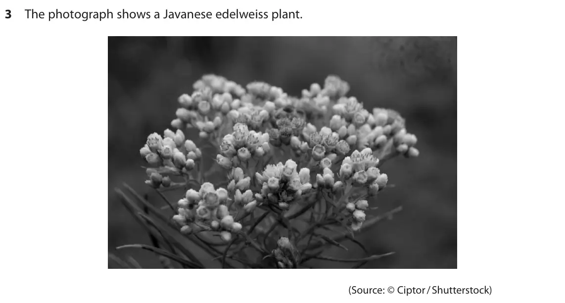 Your answer
Q1(a)(i)
The whole paper ↗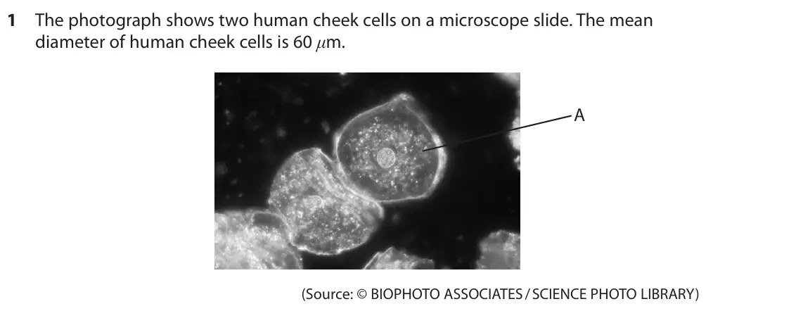
Q1(a)(ii)
The whole paper ↗Q1(b)(ii)
The whole paper ↗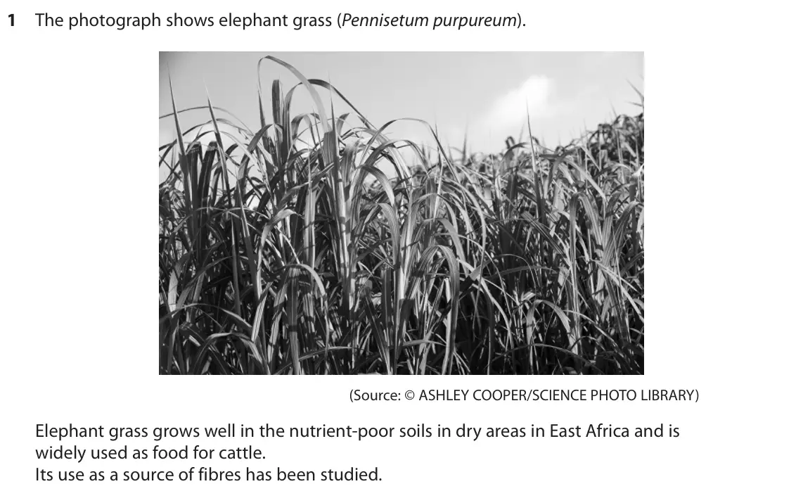
Q1(a)(iii)
The whole paper ↗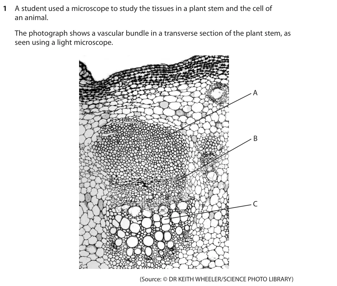
Q1(a)(iv)
The whole paper ↗Q1(b)(i)
The whole paper ↗Q2(a)(i)
The whole paper ↗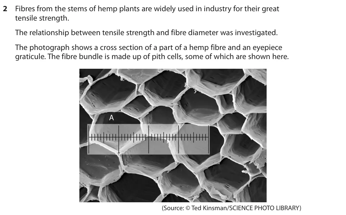
Q1(a)
The whole paper ↗
Q1(b)(i)
The whole paper ↗Q1(b)(ii)
The whole paper ↗Q3(a)(i)
The whole paper ↗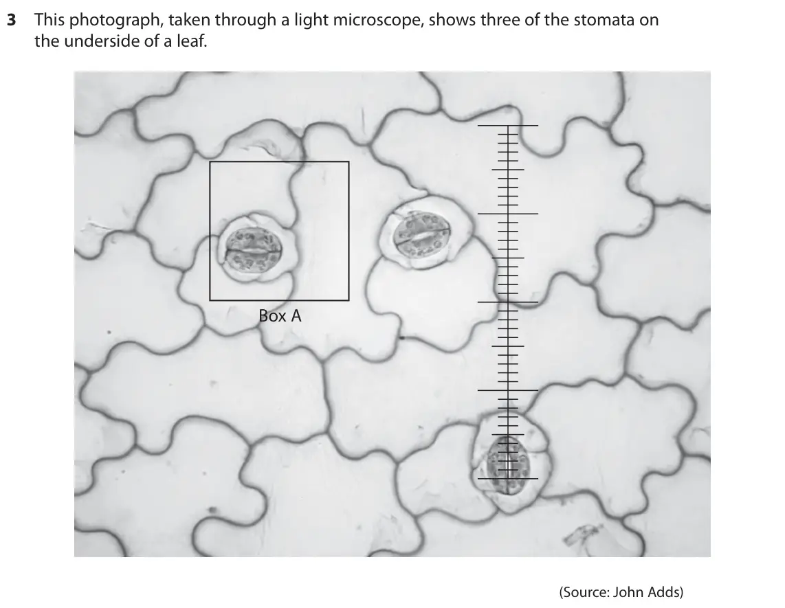
Q3(a)(ii)
The whole paper ↗Q2(b)(i)
The whole paper ↗
All 14 questions on Using a light microscope and graticule, as a list
- October 2024, AS Unit 2, Q3(b)(i) (1 mark): Which is the length of this pollen grain? (1) A 2.3 mm B 23 µm C 230 µm D 2300 nm
- October 2023, AS Unit 3, Q1(a)(i) (5 marks): Describe a procedure for the accurate determination of the diameter of cell A. (5)
- October 2023, AS Unit 3, Q1(a)(ii) (4 marks): Draw cell A and label the parts of the cell. Include a scale by your drawing. (4)
- June 2023, AS Unit 3, Q1(b)(ii) (3 marks): After soaking, the diameter of each fibre was measured. Describe how fibre diameter could be measured accurately using a microscope. (3)
- January 2023, AS Unit 3, Q1(a)(iii) (2 marks): This photograph shows a high-power view of some of the cells in tissue C in the vascular bundle. Each of the smallest units on the eyepiece graticule shown in t
- January 2023, AS Unit 3, Q1(a)(iv) (1 mark): A cell in tissue B of the vascular bundle has a cross-sectional area of 6.13 × 10⁻¹¹ m2. Calculate how many times larger the cross-sectional area of cell F is t
- January 2023, AS Unit 3, Q1(b)(i) (3 marks): The photograph shows an animal cell. This photograph was taken using an electron microscope. (Source: © DR GOPAL MURTI/SCIENCE PHOTO LIBRARY) Draw this cell as
- January 2022, AS Unit 3, Q2(a)(i) (2 marks): Each of the smallest units on the graticule is 3 × 10⁻⁶ m. Calculate the width of the pith cell labeled A in micrometres (μm). (2) Answer μm
- January 2021, AS Unit 3, Q1(a) (4 marks): Describe a safe method to prepare and examine the structure of human cheek cells. (4)
- January 2021, AS Unit 3, Q1(b)(i) (2 marks): Draw the cell labeled W in the photograph. Label two structures on your drawing. (2)
- January 2021, AS Unit 3, Q1(b)(ii) (2 marks): The photograph shows an eyepiece graticule over the widest part of this human cheek cell. (Source: © BIOPHOTO ASSOCIATES/SCIENCE PHOTO LIBRARY) Each of the smal
- October 2020, AS Unit 3, Q3(a)(i) (3 marks): Draw the cells within Box A on the photograph. (3)
- October 2020, AS Unit 3, Q3(a)(ii) (2 marks): An eyepiece graticule is shown over one of the pores. Each of the smallest units on the graticule is 3 × 10⁻⁶ m. Calculate the length of this pore in micrometre
- January 2020, AS Unit 3, Q2(b)(i) (5 marks): Devise a method to determine the cross-sectional area of a fibre, using the following equipment: • a sharp blade • a microscope • a microscope slide and coversl
Questions, mark schemes and examiner’s reports are © Pearson Edexcel, from their published past papers. Plumi isn’t affiliated with any exam board. Want whole papers? Edexcel International A Level Biology past papers.
The Plumi iPad app also offers exam-style questions with AI feedback on your handwritten working.