 plumi
Ultrasound imaging: AQA A‑Level Physics questions
AQA A-Level Physics past-paper questions on 3.10.4.1 Ultrasound imaging, 2008–2025, with mark schemes and examiner’s reports where available.
Paper
All papers
Exam
All years
Calculations
Q02.3
The whole paper ↗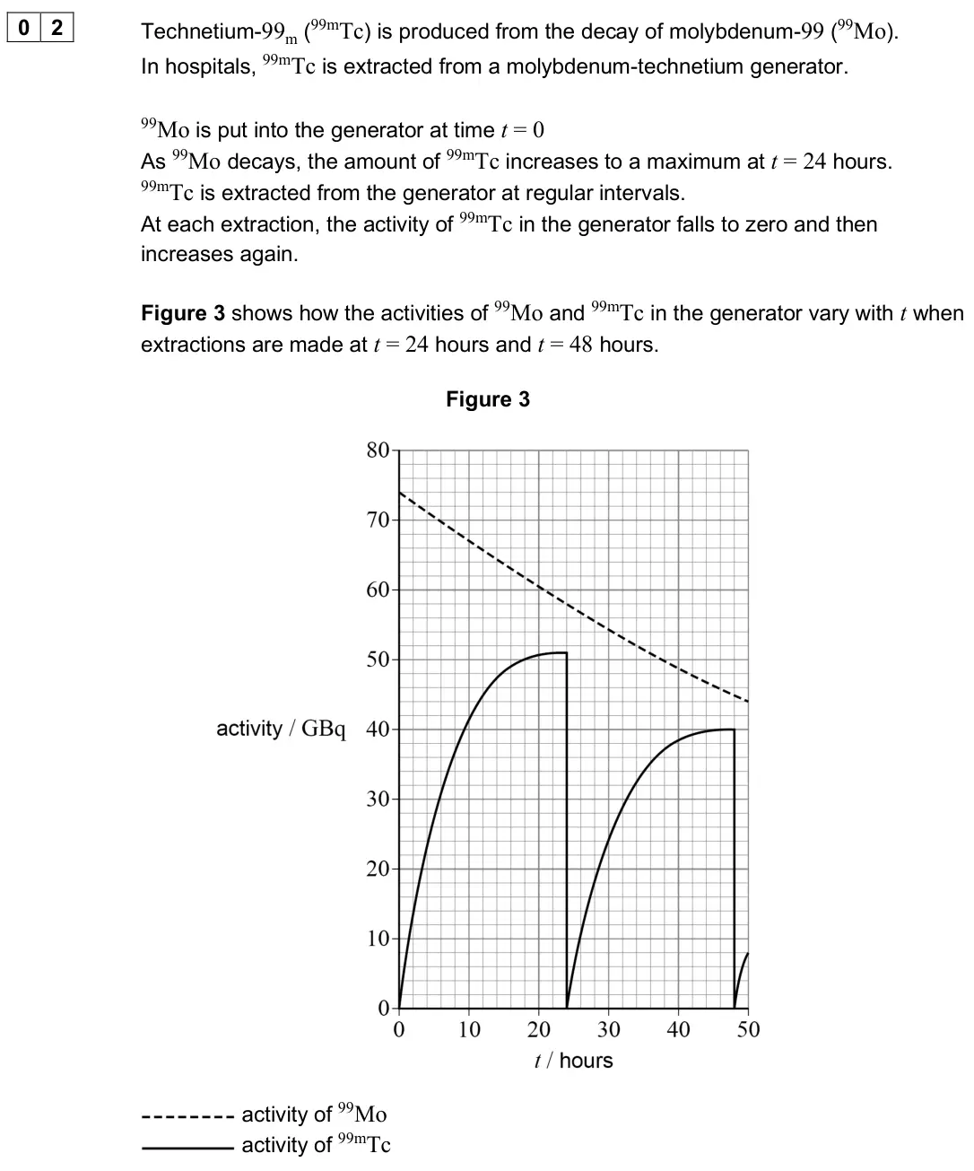
Q05.1
The whole paper ↗Q05.2
The whole paper ↗Q04.1
The whole paper ↗
Q04.2
The whole paper ↗Q04.1
The whole paper ↗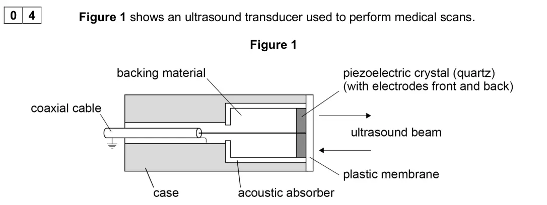
Q04.2
The whole paper ↗Q04.3
The whole paper ↗Q04.4
The whole paper ↗Q04.1
The whole paper ↗
Q04.2
The whole paper ↗Q02.2
The whole paper ↗Q02.3
The whole paper ↗Q3(a)
The whole paper ↗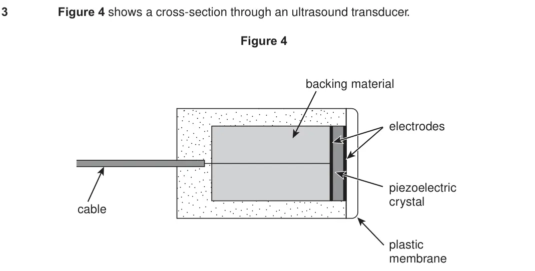
Q3(b)
The whole paper ↗Q3(c)
The whole paper ↗Q5(b)
The whole paper ↗Q3(a)
The whole paper ↗
All 27 questions on Ultrasound imaging, as a list
- June 2025, Paper 3 Medical physics, Q02.3 (6 marks): Information about heart function can be obtained from a multi-gated acquisition (MUGA) scan or from a transthoracic echocardiogram (TTE) scan. Heart specialists
- June 2024, Paper 3 Medical physics, Q05.1 (2 marks): Figure 2 shows a transducer used in a medical ultrasound scanner. Figure 2 Explain why a backing material is used. [2 marks]
- June 2024, Paper 3 Medical physics, Q05.2 (3 marks): A beam of ultrasound is transmitted from muscle into bone. Calculate the percentage of the incident intensity that is transmitted. acoustic impedance of bone =
- June 2023, Paper 3 Medical physics, Q04.1 (1 mark): Figure 3 shows the results of an ultrasound scan of a kidney using one technique. Figure 3 Identify the type of ultrasound scan used to produce Figure 3. Explai
- June 2023, Paper 3 Medical physics, Q04.2 (3 marks): Determine the patient’s heart rate in beats per minute. [3 marks] heart rate = beats per minute
- June 2022, Paper 3 Medical physics, Q04 (6 marks): A patient has calcium kidney stones. Three types of scan are available to investigate the condition: • a magnetic resonance (MR) scan • a CT scan • an ultrasoun
- November 2020, Paper 3 Medical physics, Q04.1 (6 marks): Explain how the transducer in Figure 1 operates in medical diagnosis. In your answer you should explain how • an ultrasound pulse is produced by the transducer
- November 2020, Paper 3 Medical physics, Q04.2 (1 mark): Ultrasound of frequency 1.0 MHz is used to scan a person’s liver. Estimate the resolution of the scan. speed of sound in liver tissue = 1600 m s−1 [1 mark] reso
- November 2020, Paper 3 Medical physics, Q04.3 (4 marks): Ultrasound travels from a transducer through the chest wall to an air pocket inside the lung. From the air pocket, the ultrasound is then incident on lung tissu
- November 2020, Paper 3 Medical physics, Q04.4 (2 marks): Discuss whether an ultrasound scan would be suitable to investigate a tumour inside a lung. [2 marks]
- June 2019, Paper 3 Medical physics, Q04.1 (3 marks): An ultrasound A-scan is used to find the length l of an eye as shown in Figure 6. Figure 7 shows the simplified A-scan for the eye. A short pulse of ultrasound
- June 2019, Paper 3 Medical physics, Q04.2 (6 marks): Amniocentesis is a procedure where a tube is inserted into a uterus to remove some cells and fluid from around a foetus. For the procedure to be carried out saf
- June 2017, Paper 3 Medical physics, Q02.2 (2 marks): The blood vessel called the aorta passes through the abdomen. A second patient with a suspected fault in the wall of the aorta can be given an ultrasound scan o
- June 2017, Paper 3 Medical physics, Q02.3 (4 marks): When ultrasound travels across a boundary from blood to the wall of the aorta there is a decrease in acoustic impedance across the boundary. This results in 0.0
- Specimen, Paper 3 Medical physics, Q03 (6 marks): Positron Emission Tomography (PET) and ultrasound scans are both used in medica diagnosis. Compare the quality of the information obtained from these scans in t
- June 2016, PHYA5 Medical physics (old spec), Q3(a) (3 marks): Explain how the transducer produces a pulse of ultrasound. [3 marks] ______________________________________________________________________________ ____________
- June 2016, PHYA5 Medical physics (old spec), Q3(b) (1 mark): Why does the ultrasound pulse produced need to be short? Place a tick () in the right-hand column to show the correct answer. [1 mark] if correct To reduce pu
- June 2016, PHYA5 Medical physics (old spec), Q3(c) (3 marks): A coupling gel is needed when performing an ultrasound scan. Explain how and why a coupling gel is used. [3 marks] _____________________________________________
- June 2014, PHYA5 Medical physics (old spec), Q5(b) (2 marks): Ultrasound is thought to be more suitable than X-rays for scanning an unborn fetus. State two reasons why ultrasound is used for this application. [2 marks] rea
- June 2013, PHYA5 Medical physics (old spec), Q3(a) (4 marks): Figure 1 shows an ultrasound transducer used in an A-scan. Figure 1 Outline, with reference to the diagram, the process by which the transducer produces a short
- June 2013, PHYA5 Medical physics (old spec), Q3(b)(i) (2 marks): Calculate the percentage of the incident ultrasound which would be transmitted into the skin when incident on an air-skin boundary. acoustic impedance of air =
- June 2013, PHYA5 Medical physics (old spec), Q3(b)(ii) (2 marks): When obtaining the ultrasound image of an unborn foetus, a coupling gel is used. Explain why a coupling gel is needed and state the property of the gel that ens
- June 2012, PHYA5 Medical physics (old spec), Q4(a) (6 marks): Explain how and why ultrasound is used to obtain an image of an unborn foetus. You might consider the following points in your answer l the method of obtaining
- June 2012, PHYA5 Medical physics (old spec), Q4(b) (2 marks): Explain why the pulses of ultrasound used in medical imaging must be of short duration.
- June 2008, PHA6W (2008 spec), Q4(a) (2 marks): 4 (a) Figure 1 shows a cross-section through an ultrasound generator. 4 (a) Explain the purpose of the backing material. (2 marks) Figure 1 backing material ele
- June 2008, PHA6W (2008 spec), Q4(b) (1 mark): 4 (b) State the main difference between the probe used in an A-scan and that used in a B-scan. (1 mark)
- June 2008, PHA6W (2008 spec), Q4(c) (4 marks): 4 (c) Figure 2 shows the display on an oscilloscope screen of an A-scan to find the size of an organ. Figure 2 strength of signal generated pulse spinal column
Questions, mark schemes and examiner’s reports are © AQA, from their published past papers. Plumi isn’t affiliated with any exam board. Want whole papers? AQA A-Level Physics past papers.
The Plumi iPad app also offers exam-style questions with AI feedback on your handwritten working.