 plumi
The physics of diagnostic X-rays: AQA A‑Level Physics questions
AQA A-Level Physics past-paper questions on 3.10.5.1 The physics of diagnostic X-rays, 2008–2025, with mark schemes and examiner’s reports where available.
Paper
All papers
Exam
All years
Calculations
Q04.1
The whole paper ↗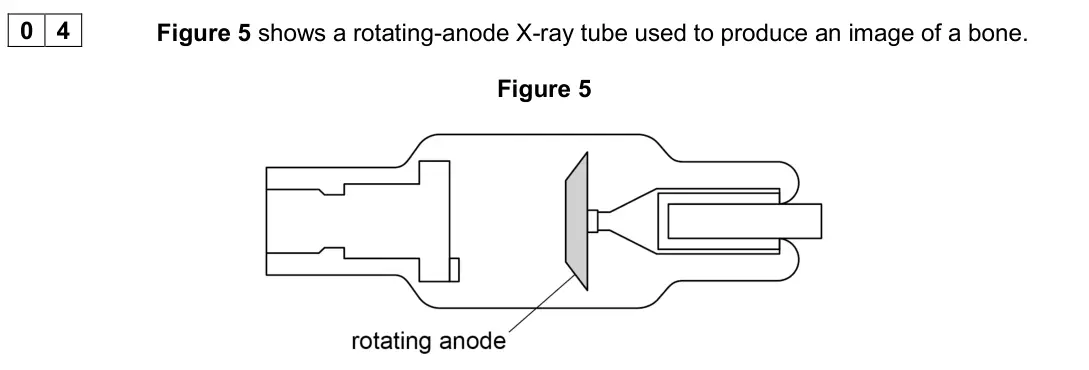
Q04.2
The whole paper ↗Q04.1
The whole paper ↗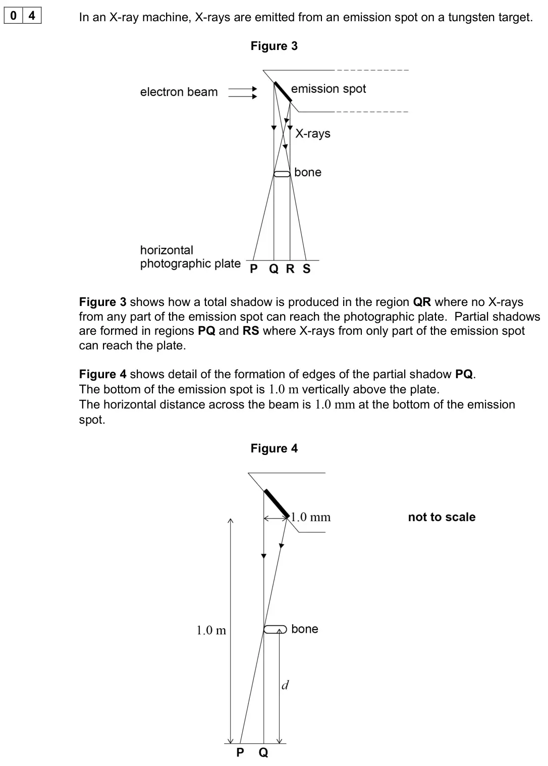
Q04.2
The whole paper ↗Q03.1
The whole paper ↗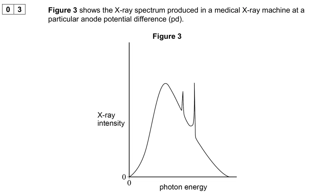
Q03.2
The whole paper ↗Q03.3
The whole paper ↗Q02.1
The whole paper ↗Q4(a)
The whole paper ↗Q4(a)(i)
The whole paper ↗
Q4(a)(ii)
The whole paper ↗Q5(a)(i)
The whole paper ↗
Q5(a)(ii)
The whole paper ↗Q5(a)
The whole paper ↗Q5(a)
The whole paper ↗Q5(b)
The whole paper ↗
All 17 questions on The physics of diagnostic X-rays, as a list
- June 2025, Paper 3 Medical physics, Q04.1 (2 marks): X-rays originate from a region on the rotating anode known as the focal spot. Explain: • one advantage of using a small focal spot • why the focal spot should n
- June 2025, Paper 3 Medical physics, Q04.2 (3 marks): State and explain three methods used to limit exposure of the patient’s healthy cells to X-rays during a diagnostic procedure. [3 marks] 1 2 3
- November 2021, Paper 3 Medical physics, Q04.1 (2 marks): To produce a sharp image of a bone, the partial shadow in region PQ must be no more than 0.10 mm wide. Calculate the maximum distance d between a bone and the p
- November 2021, Paper 3 Medical physics, Q04.2 (2 marks): Discuss whether an X-ray image of a chest or an X-ray image of a hand is likely to be sharper when exposed to the same X-ray source. [2 marks]
- June 2019, Paper 3 Medical physics, Q03.1 (4 marks): In an X-ray tube, electrons collide with a tungsten target. Explain how the continuous spectrum and the characteristic spectra are produced by these electron co
- June 2019, Paper 3 Medical physics, Q03.2 (2 marks): The dashed line on Figure 4 shows the X-ray spectrum for the initial anode pd. Sketch on Figure 4 the X-ray spectrum produced when the anode pd is increased. [2
- June 2019, Paper 3 Medical physics, Q03.3 (1 mark): In the medical X-ray machine, the X-rays produced with the initial anode pd are now passed through an aluminium filter. The dashed line on Figure 5 shows the X-
- June 2018, Paper 3 Medical physics, Q03 (6 marks): In the past, doctors could only use a simple X-ray image to assess head injuries. A CT scan is now a preferred technique. Discuss why the CT scan has replaced t
- June 2017, Paper 3 Medical physics, Q02.1 (6 marks): A patient with a suspected broken arm is going to have an X-ray image taken. Explain the risk to the patient of exposure to X-rays. Go on to discuss three ways
- June 2016, PHYA5 Medical physics (old spec), Q4(a) (6 marks): Figure 5 shows a simplified modern X-ray tube with a rotating anode. Figure 5 Explain the design and operation of the X-ray tube and the purposes of the compone
- June 2014, PHYA5 Medical physics (old spec), Q4(a)(i) (1 mark): State the use of the lead sheets. [1 mark]
- June 2014, PHYA5 Medical physics (old spec), Q4(a)(ii) (1 mark): State the use of the aluminium filter. [1 mark]
- June 2012, PHYA5 Medical physics (old spec), Q5(a)(i) (2 marks): Calculate the kinetic energy of an electron as it reaches the anode. Give your answer to an appropriate number of significant figures. answer = J (2 marks)
- June 2012, PHYA5 Medical physics (old spec), Q5(a)(ii) (2 marks): Assuming that the electron gives up all this energy to form an X-ray photon, calculate the wavelength of the photon. answer = m (2 marks)
- June 2011, PHYA5 Medical physics (old spec), Q5(a) (3 marks): The X-ray spectrum for a certain X-ray tube target is shown in Figure 4. Explain the process which gives rise to spikes at certain photon energies. Figure 4
- June 2008, PHA6W (2008 spec), Q5(a) (2 marks): 5 (a) A rotating anode X-ray tube is operated at a peak voltage of 90 kV and an electron beam current of 48 mA. 5 (a) (i) Calculate the number of electrons reac
- June 2008, PHA6W (2008 spec), Q5(b) (4 marks): 5 (b) State and explain two ways to reduce the radiation energy absorbed by the patient when obtaining an X-ray photographic image of good quality. First way Se
Questions, mark schemes and examiner’s reports are © AQA, from their published past papers. Plumi isn’t affiliated with any exam board. Want whole papers? AQA A-Level Physics past papers.
The Plumi iPad app also offers exam-style questions with AI feedback on your handwritten working.